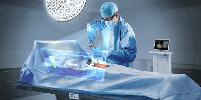
A new study from the School of Medicine, Department of Neurosurgery, Technical University of Munich, Germany evaluated an augmented reality (AR) navigation system for pedicle screw placement while integrating navigation cameras into the reference array and drill guide.
How Accurate Is Screw Placement Using AR?
3 min read Premium comments
#spinesurgerySecondary#pediclescrewplacement#augmentedreality
Lead author, Professor Dr. med. Bernhard Meyer, Director of the Neurosurgical Clinic and Policlinic at the University Hospital Rechts der Isar, Munich summarized his findings, saying: “Precision and accuracy in spine surgery is critical. As stated in our recent study, AR-assisted navigation greatly simplified pedicle screw placement, ensuring effectiveness and accuracy. It’s a reliable tool, straightforward to use, and it elevates our ability to provide safe and precise navigation for various spinal conditions.”
The study was published in the October 16, 2023 edition of the European Spine Journal: “Evaluating a cutting‑edge augmented reality‑supported navigation system for spinal instrumentation”1
The goal of the new study was to evaluate the safety, efficacy, and accuracy of an augmented reality surgery navigation system on a total of 20 patients who were operated on between June 2021 and January 2022. Of the 20 patients, 10 were women and 10 were men, average age 69.4 years (range was 32.1 – 88.0).
The system (NextAR from Medacta, Castel San Pietro, Switzerland) relies on a new technique for intraoperative navigation. The research team evaluated the system’s accuracy of pedicle screw placement using intraoperative CT imaging.
- A median of 8 (4–18) pedicle screws were placed in each case.
- Percutaneous instrumentation was performed in 14 patients (70%).
- The duration of pedicle screw placement (duration scan–scan) was 56±26 (30–107) min.
- Intraoperative screw revision was necessary for 3 of 180 pedicle screws (1.7%).
Intraoperatively, no major complications occurred—one case of delay due to software issues and one case of difficult screw placement were reported.
Specifically, the authors found that in either open or percutaneous approaches in both long and short constructs, using the augmented reality navigation system resulted in a 1.7% intraoperative revision rate as compared to the rate documented in the literature of 3.4% by Ille at al2 to 4.7% by Ryang at al3.
The Surgery Itself
The surgeries were either an open midline approach or percutaneous pedicle screw implantation depending on the additional need for decompression, construct length, and instrumentation levels.
For spinal navigation, the surgical team attached a reference array equipped with navigation targets incorporating multiple light emitting diodes (LEDs) in a certain spatial configuration to a spinous process close to the levels planned for instrumentation, and an infrared camera was also firmly attached to surgical instruments such as the drill guide.
Next, the team performed a navigation scan using either a 3D C-arm scanner (Vision, Ziehm Imaging, Nuremberg, Germany) or an operation room-based CT scanner (ORCT) (Brilliance CT Big Bore, Philips, Amsterdam, Netherlands).
After uploading the navigation data set to the navigation software, the surgical team reviewed the image quality, as well as the accuracy of the registration in correlation with anatomical structures.
The surgical team then deployed the AR-supported navigation system (NextAR from Medacta, Medacta, Castel San Pietro, Switzerland), performed skin incisions for percutaneous instrumentation, screw entry points, and panned screw trajectories using a navigated drill guide.
Once planned, the surgical team followed trajectories using a battery-powered drill. They inserted a Kirschner wire via the drill guide to mark the screw trajectory. Next, the surgeon implanted cannulated pedicle screws (Medacta Universal Screw Technology [MUST], Medacta, Castel San Pietro, Switzerland) by passing them over the Kirschner wires.
Once the pedicle screws were in place, the team acquired a final control scan and then reviewed pedicle screw placement on 3D imaging.
In case of screw revision, the team performed another final scan. Afterward, the team fitted rods according to curvature and length and installed them by fixating the pedicle screw nuts.
Finally, the team performed additional decompression depending on the surgical indication of surgery.
Dr. Meyer and his team received no funding from Medacta or any other suppliers of implants or instruments or systems used in the surgeries.
REFERENCES
[1] Schwendner M, Ille S, Wostrack M, Meyer B. Evaluating a cutting-edge augmented reality-supported navigation system for spinal instrumentation. Eur Spine J. 2023 Nov 14. doi: 10.1007/s00586-023-08011-w. Epub ahead of print. PMID: 37962688.
[2] Ille S, Baumgart L, Obermueller T, Meyer B, Krieg SM (2021) Clinical efficiency of operating room-based sliding gantry CT as compared to mobile cone-beam CT-based navigated pedicle screw placement in 853 patients and 6733 screws. Eur Spine J 30(12):3720–3730. https:// doi. org/ 10. 1007/ s00586- 021- 06981-3
[3] Ryang YM, Villard J, Obermuller T et al (2015) Learning curve of 3D fluoroscopy image-guided pedicle screw placement in the thoracolumbar spine. Spine J 15(3):467–476. https:// doi. org/ 10. 1016/j. spinee. 2014. 10. 003
React:
Discussion
This is a fascinating development. In my practice we've seen similar outcomes with the revised protocol. The key differentiator seems to be patient selection criteria. Has anyone else noticed the correlation with BMI thresholds?
Great point. I'd push back slightly on the conclusion, the sample size in the cited study is too small to draw population-level inferences. That said, the directional signal is compelling and worth a larger RCT.
We implemented a similar approach last year. Early results are promising but we're still gathering 12-month follow-up data. Happy to share our protocol if anyone is interested.
Join the conversation
Orthopedic professionals are discussing this. Sign in and upgrade to read every comment and add your voice.