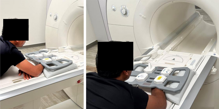
Patient having his wrist scanned with a video producing low power MRI / Source: UC Davis
#mri#wristpain#wristsurgery
A UC Davis and Stanford team, that has been working on the problem of combining motion and MRI for ten years, has created a video of articulating wrist joint using an MRI machine. Yes, the patient moves while inside an MRI core.
Kudos to Abhijit Chaudhari, professor in the UC Davis Department of Radiology and interim director of the UC Davis Imaging Research Center; Krishna Nayak, who directs the Dynamic Imaging Science Center at the University of Southern California; Robert Szabo, distinguished professor of orthopaedics and chief emeritus of hand and upper extremity surgery at UC Davis; and Robert Boutin, professor of radiology at Stanford University and musculoskeletal radiologist.
First, they used a low-field, 0.55 tesla, MRI (magnetic resonance imaging) system. Standard MRI’s use 1.5 – 3.0 tesla power. In this instance the word “tesla” is the scientific unit of measure for magnetic strength. A refrigerator magnet, for example, uses roughly 0.001 tesla power output. A standard MRI, therefore, would be using the equivalent of 1.5-3.0 million refrigerator magnets to generate soft tissue images.
Also, of course, this unit of measure honors one of the greatest inventors and electrical engineers in history—Nikola Tesla, who died in 1943.
But we digress. How did Chaudhari and Boutin combine motion with MRI imaging?
From their study, the two authors described how a novel low power MRI was able to capture wrist motion:
Advertisement
“High-performance 0.55T systems offer a high acquisition duty cycle, typically achieved using efficient k-space trajectories, not possible or practical at higher field strengths and resulting in the mitigation of image artifacts. These systems can further utilize pulse sequences, such as balanced steady-state press precession (bSSFP), that provide superior signal-to-noise ratio and efficiency enabling a vast increase in imaging speed compared to higher field system.”
So, in effect, using a high-performance, but much lower power MRI produced cleaner images, at a higher acquisition duty cycle. More and better images faster—fast enough, in fact, to create a video of wrist joint motion.
Anyone who’s had an MRI remembers the high-power MRI machines clunk along, slowly taking one picture, then another—while being told to lies motionless for several minutes.
We asked Drs. Sabo and Chaudhari about the incongruence of using MRI to capture motion videos of articulating joints and they said: “Indeed, typically patients are asked to lie motionless on the scanner. However, several pathological conditions underlying wrist dysfunction occur only during motion or when performing specific tasks using our hands.”
“These conditions often result in pain, limited range-of-motion and loss of strength. Therefore, there was a strong motivation to ‘look inside’ the wrist and understand these conditions when the wrist is moving.”
“Higher field strengths (1.5T or 3.0T) provide outstanding image quality and allow us to observe minute changes in tissues of the stationary wrist. However, when motion is conducted, that disturbs the high magnetic field, and the resulting images show severe artifacts.”
“At low field (0.55T), there is much less perturbation of the magnetic field, given that the field itself is low. Therefore, images during motion demonstrate much lower artifacts. This helps in visualizing the wrist tissues during motion and assess the trajectories of the various tissues.”
“An obvious question is that while images during motion are better quality using 0.55T, what about images of the static wrist? Are they reasonable quality so that we can use images of the stationary and the moving wrist together for assessing wrist dysfunction? Our paper demonstrates that this indeed is the case, and therefore a comprehensive assessment of wrist dysfunction should be possible with the 0.55T system.”
Advertisement
What’s next?
As has been established by many other studies, imaging articulating joints under loads and/or in motion, can change the game when it comes to diagnosing soft tissue injuries.
So, of course, we asked the study authors what they thought were next steps for them. They told OTW:
“Our next steps include several studies:
To use this technique to examine patients with wrist pain and normal X-rays and normal MRIs and see if there is any pathology that can be identified.
To study different normal wrists which have different motion patterns established with static images and see how these structures move in real time to understand how factors such as laxity relates to instability.
To study abnormal wrists with known ligament tears in either the carpal or distal radio-ulnar joints and demonstrate how that affects motion.
To compare preoperative and postoperative studies early and long-term to evaluate the success of different wrist reconstructions for carpal instability problems.
And that is just the beginning!!”
The wrist has eight carpal bones and many ligaments. It is not uncommon for patients with chronic wrist pain to be told, after their doctor takes several static MRI or CT images, that their pain is indeterminant.
The imaging didn’t find the problem.
“Moving images give us a new tool to diagnose wrist dysfunction, either during motion or when there is load on the joint,” said Abhijit Chaudhari. “The wrist is highly complex, so the ability to visualize motion will have enormous impact.”
Advertisement
Ten Year Project, So Far
For Chaudhari, Szabo and Boutin this work started ten years ago, in 2013. Initially, they could only produce two to three MRI frames per second—using, notably, a powerful three tesla (3T) MRI machine. The 3T machine produced visual artifacts, such as bands without signal, which obscured wrist anatomy.
“We were doing all our work on 3T systems, and we realized we could not do much better because these systems have limitations,” said Chaudhari. “Whenever the patient moved within the high-field strength magnet, they disturbed the magnetic field a lot, and that created artifacts in images that disrupted our ability to assess the joints.”
Developing the 0.55T System
Krishna Nayak, who directs the Dynamic Imaging Science Center at the University of Southern California and is a co-author on the new paper, had received a National Science Foundation grant to develop a 0.55T MRI system specifically for dynamic imaging.
Chaudhari, Szabo and Boutin then teamed up with Nayak to see if his 0.55T machine could produce diagnostic-quality images, either as stills or as a short, 78 frame-per-second movie.
Nayak’s 0.55T machine was incredibly fast. It produced high-quality videos in 5 seconds or less.
The researchers discovered that Nayak’s rapid capture MRI delivered videos without adding significant cost.
Advertisement
“This approach allows us to study the trajectory of structures in an actively moving wrist,” said Szabo. “The dynamic pictures, along with standard, still MRI scans, show us specific wrist anatomy that had never been evaluated to this degree before. This has tremendous relevance to evaluate injuries and conduct further research into how the wrist functions.”
A Wealth of Applications
In addition to advancing real-time MRI, the new 0.55T system could provide much-needed clinical versatility. The magnetic field does not extend far beyond the machine, eliminating the risk of disturbing metallic devices in the room. The instrument could also help patients with surgical implants, who currently cannot use high-field MRIs. Also, the machine has a smaller footprint, allowing it to be placed in smaller rooms.
“This could be quite beneficial for intraoperative applications, in which you take the patient out of the scanner and immediately perform the intervention,” said Chaudhari. “The applications of these low-field scanners continue to expand.”
“Using this technique before and after wrist reconstructive surgery would allow us to see if our techniques restore normal anatomy in our patients,” said Szabo. “Imagine the difference between taking a movie of someone’s wrist while they are throwing a baseball compared to taking a still picture. Now imagine taking a moving MRI showing the inside of the wrist compared to an X-ray or static MRI. The added information will really improve care.”
Additional co-authors on the paper include Christopher Bayne of UC Davis, Yongwan Lim of the University of Southern California, and Sophia X. Cui of Siemens Medical Solutions.
The study sponsors were the National Science Foundation Grant MRI-1828736 and Siemens Healthineers.
" data-large-file="https://i0.wp.com/ryortho.com/wp-content/uploads/2023/10/MRI_WristMRI_WEB.jpg?fit=850%2C253&ssl=1" src="https://i0.wp.com/ryortho.com/wp-content/uploads/2023/10/MRI_WristMRI_WEB.jpg?resize=850%2C253&ssl=1" alt="" width="850" height="253">Here are some static images of the wrist in the coronal plane ; (a) proton-density weighted 2D fat-suppressed, TSE image showing the visualization of the SL ligament (orange arrow) and the TFCC (green arrow); (b) T1w 2D TSE image demonstrating the anatomical structures of the wrist; and (c) 3D T1w gradient recalled echo pulse sequence with Dixon-based water reconstruction, illustrating the anatomical template to segment the bones. / Source: UC Davis
" data-large-file="https://i0.wp.com/ryortho.com/wp-content/uploads/2023/10/MRI_WristMRI2_WEB.jpg?fit=850%2C665&ssl=1" src="https://i0.wp.com/ryortho.com/wp-content/uploads/2023/10/MRI_WristMRI2_WEB.jpg?resize=850%2C665&ssl=1" alt="" width="850" height="665">Here are images of the wrist under motion, performing the RUD maneuver with varying temporal resolution, with a spiral bSSFP sequence. Snapshots of the wrist in the neutral position with full sampling (164.6 ms per image), and with acceleration by factors of 92%, 85 and 77%. Temporal blurring is induced by lower temporal resolution and obscures the visualization of the wrist joints (red arrow shows the SL interval). Slice thickness is 6 mm. / Source: UC Davis
React:
Discussion
14
DS
Dr. Sarah MitchellOrthopedic Surgeon · Mayo Clinic
This is a fascinating development. In my practice we've seen similar outcomes with the revised protocol. The key differentiator seems to be patient selection criteria. Has anyone else noticed the correlation with BMI thresholds?
8
JT
James Thornton, MDSpine Fellow · HSS
Great point. I'd push back slightly on the conclusion, the sample size in the cited study is too small to draw population-level inferences. That said, the directional signal is compelling and worth a larger RCT.
5
RP
R. PatelSports Medicine · Stanford
We implemented a similar approach last year. Early results are promising but we're still gathering 12-month follow-up data. Happy to share our protocol if anyone is interested.
Join the conversation
Orthopedic professionals are discussing this. Sign in and upgrade to read every comment and add your voice.
Discussion
This is a fascinating development. In my practice we've seen similar outcomes with the revised protocol. The key differentiator seems to be patient selection criteria. Has anyone else noticed the correlation with BMI thresholds?
Great point. I'd push back slightly on the conclusion, the sample size in the cited study is too small to draw population-level inferences. That said, the directional signal is compelling and worth a larger RCT.
We implemented a similar approach last year. Early results are promising but we're still gathering 12-month follow-up data. Happy to share our protocol if anyone is interested.
Join the conversation
Orthopedic professionals are discussing this. Sign in and upgrade to read every comment and add your voice.