What could be more patriotic than some biologic fireworks courtesy of the National Institutes of Health and one of their NIH funded teams?
Simply the Coolest Biologic Video Ever
1 min read Premium comments
Secondary
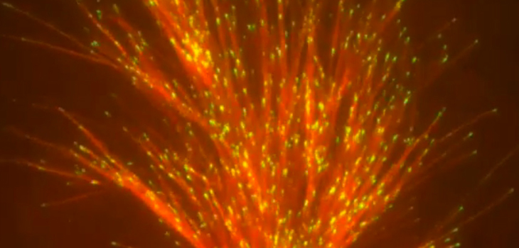
Hats off to the kick-ass team led by Sabine Petry, Ph.D. at Princeton University, Princeton, New Jersey. As part of their study of the dynamics of microtubules, the researchers created a time-lapse video of fluorescence-tagged proteins. Using a specialized microscope equipped with a time lapse camera this NIH-funded team captured a critical step in the process of cell division, or mitosis. They showed how filaments called microtubules (red) form new branches (green) and fan out to form mitotic spindles.
The team added pyrotechnic sound effects. And created, truly, the coolest biologic video ever!
Sabine Petry’s team is studying the dynamics of microtubules in a cell-free extract of cytoplasm taken from the egg of an African clawed frog (Xenopus laevis). Petry’s ultimate goal is to learn how to build mitotic spindles, molecule by molecule, in the lab. Such an achievement would mark a major step forward in understanding the complicated mechanics of cell division, which, when disrupted, can cause cancer and many other health problems.
React:
Discussion
This is a fascinating development. In my practice we've seen similar outcomes with the revised protocol. The key differentiator seems to be patient selection criteria. Has anyone else noticed the correlation with BMI thresholds?
Great point. I'd push back slightly on the conclusion, the sample size in the cited study is too small to draw population-level inferences. That said, the directional signal is compelling and worth a larger RCT.
We implemented a similar approach last year. Early results are promising but we're still gathering 12-month follow-up data. Happy to share our protocol if anyone is interested.
Join the conversation
Orthopedic professionals are discussing this. Sign in and upgrade to read every comment and add your voice.