If you can think of it, these guys can make it—right now. Give them your dreams, your X-rays, your MRIs, your sketches on napkins and they will give you back an implant, an instrument or pretty much anything you’ve imagined. In short, they can plug your mind into a fabrication machine.
From Your Lips to the Machine’s Ear
6 min read Premium comments
Welcome to a revolution called e-manufacturing. One small Connecticut-based company has just made a big bet on this concept. The company, Oxford Performance Materials, LLC (OPM), began a multi-million dollar program earlier this year to, literally, take anything a surgeon or engineer dreams of: X-rays, MRIs or napkin sketches, and produce exactly that part—right now.
Plugging Your Mind Into the Machine
The e-manufacturing process that OPM is building has been revolutionizing the aircraft, auto and other manufacturing industries and is now about to (FDA willing and the CMS don’t rise) accelerate innovation and time to market in orthopedics.
E-manufacturing is the system for taking any kind of digital input from any source and then sending it directly to a machine via the Internet (or Intranet). E-manufacturing effectively makes the thoughts of an inventor transparent to the fabricating machine.
For years, the electronics of e-manufacturing have been running far ahead of the fabricating machines. Up until just recently, e-manufacturing machines created prototype products from low temperature, low performance polymers. Then a German company named EOS presenting at the Euromold Plastics Convention introduced a digital laser sintering machine (like a 3D laser printer) which could melt high performance polymeric powders and fuse them into solid shapes. We’re talking melting points as high as 380°C (716°F) degrees. This was a huge innovation. With the EOS printer, e-manufacturing systems could now move to high performance, medical grade polymers like PEKK.
It took one full year to sell the first machine. That was two years ago.
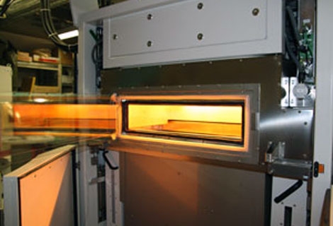
 Mastoid form created through the
OXPEKK OsteoFab manufacturing
process. Source: Oxford
Performance Materials, LLCEven now, there are barely a handful of these machines in use around the world—including, of course, the one operating at 30 South Satellite Road in South Windsor, Connecticut (OPM’s corporate headquarters).
OPM’s system is in fact the world’s first system that can plug into an e-manufacturing system and laser-sinter PEKK polymers. OPM can fabricate virtually any implant or instrument imaginable.
Since the OPM’s new EOS machine is also on the Internet, it can be tether-free (i.e., wireless) and can make use of advanced predictive technologies. In the jargon of manufacturing, OPM’s system pushes surgeon, engineer or inventor data (typically images) through a distributed, flexible, open, reconfigurable, scalable and extendable communication network which spits out, at the end of it all, a fabricated PEKK polymer implant or instrument.
Need for Speed
Probably e-manufacturing’s biggest revolution is its ability to create bionic structures which, up until now, would have been inconceivable using conventional manufacturing methods(try making that mastoid form in the attached image without e-manufacturing capabilities).
Here’s what implant creation was like many years ago.
(The following account comes from AAOS’s inspiring history of orthopedics book “Moving Stories: 75 Years of Orthopaedics.”
In late September 1987 a six-month-old boy arrived at Christus Santa Rosa Children’s Hospital with seemingly irreversible defects—the most severe of which was a missing left chest wall. Seven of the twelve ribs were gone, so there was no support for the lung. The child also had severe scoliosis. Doctors in Houston had tried to treat the chest wall defect with a splint, but had been unsuccessful. The child was out of options and had been sent home to die.
Robert Campbell, M.D. was the orthopedic surgeon and Melvin Smith, M.D. was the pediatric general surgeon in charge of this young boy’s case.
“I didn’t know it at the time, but I was about the third orthopod Dr. Smith had asked and the other two had said sorry, but there’s nothing to be done, ” Dr. Campbell recalls with a slight smile. While that may be true, Dr. Smith remembers he was just trying to find help from “somebody who had as wild ideas as I did.”
Dr. Smith suggested using some plates or rods that could be screwed into place, but any screws used around the chest would work loose and perhaps go into the heart. Dr. Campbell went home that night and after a bit of doodling, settled on using fracture pins—metal pins used to stabilize and realign broken bones.
But it was one thing to make some doodles but quite another to make those doodles real—inside a sick infant’s chest.

The two surgeons went to work. As Dr. Campbell recalled; “I stuck the first Steinmann pin up there, got sterile vice grips, and started bending them around the ribs of this six-month-old infant. It was just terrible because I had to use tremendous force. One slip and I would have torn the axillary artery or damaged the spinal cord…my shoes were filling up with sweat.”
Despite everything, the surgery was successful. They had saved his life. But very soon new problems emerged. The pins wouldn’t grow as the boy matured and therefore the makeshift device would end up harming the patient.
Dr. Campbell decided he needed to invent a device to replace the Steinmann pins.
He approached a large orthopedic implant manufacturer and after two months of negotiating the confidentiality agreement he moved on. The second company he approached took a month before telling him that they couldn’t do it but referred him to a custom prosthesis firm in California called Techmedica.
Dr. Campbell sketched his designs out for Techmedica on children’s construction paper. He then transferred those sketches to an actual blueprint. His invention was an expandable rib—kind of like a curtain rod for the chest.

Nearly a year after the original surgery, with the original patient starting to outgrow the Steinmann pins and with the scoliosis returning, Dr. Campbell sat down with Techmedica’s engineers and got to work. It would take another half year of trading blueprints back and forth for the company to create the first prototype.
Time was running out for his patient. On April 18, 1989 the first fabricated expandable child’s ribs arrived at Dr. Campbell’s office and the very next day he implanted his Vertical Expandable Prosthetic Titanium Rib (VEPTR)—which performed as hoped for, for his young patient.
The process with everyone working as fast as they could, took from September 1987 to April 1989—18 months.
Fast forward to mid-2011. Today, with e-manufacturing systems in place, Dr. Campbell’s options change dramatically. With the Internet, Dr. Campbell would email his patient’s X-ray or MRI as well as digital images of his sketches to, for example, OPM. Before the week was out, a PEKK 3D fabricating machine would have “printed out” the first of several prototypes of the VEPTR and then the implant itself.
Just like that.
Information Transparency
When the information residing in the head of a surgeon or inventor becomes transparent to the fabricator, a new roadmap opens up for innovation and patient care.
It cost OPM more than $1.2 million to install the centerpiece of this e-manufacturing system—the EOSINT P 800 SLS machine for fabricating medical implants and instruments. OPM secured its funding from Connecticut Innovation’s BioScience Facilities Fund in the form of a special debt instrument. The machine is but one part of a multi-million dollar project to make next generation orthopedic implants through additive fabrication using OPM’s proprietary OXPEKK materials.
OPM, which was founded in March 2000, operates out of an 18, 000 square foot facility in South Windsor, Connecticut. OPM currently sells its biomedical polymeric OXPEKK products throughout the world and their customers have received regulatory approvals from the FDA, KFDA, ANVISA, COFEPRIS, and several CE Marks: OPM is ready for business anywhere.
OPM has traditionally sold its OXPEKK products as raw materials. But with the addition of the EOSINT P 800, the company will now be able to provide value‐added manufacturing in‐house. As we’ve been describing, the new remarkable fabricating machine opens up the possibility of direct digital additive manufacturing through selective laser sintering (SLS), which means that nearly anything that can be designed can be built with the firm’s implantable polymer.
We were so taken with this new technology and what it means for orthopedics, that we called OPM’s president, Scott DeFelice, and interviewed him via Skype. Sorry about the quality of this interview, BUT please listen carefully. This enhanced capability positions the firm to lead innovation in biomedical technologies and the polymer market. OPM’s initial focus is on the production of custom cranial and maxillofacial implants which are anatomically identical implants derived directly from a CT scan or MRI.
Seriously, this is remarkable and we hope that OPM’s phones start ringing off the wall from surgeons and engineers who want to get on the e-manufacturing wave.
React:
Discussion
This is a fascinating development. In my practice we've seen similar outcomes with the revised protocol. The key differentiator seems to be patient selection criteria. Has anyone else noticed the correlation with BMI thresholds?
Great point. I'd push back slightly on the conclusion, the sample size in the cited study is too small to draw population-level inferences. That said, the directional signal is compelling and worth a larger RCT.
We implemented a similar approach last year. Early results are promising but we're still gathering 12-month follow-up data. Happy to share our protocol if anyone is interested.
Join the conversation
Orthopedic professionals are discussing this. Sign in and upgrade to read every comment and add your voice.