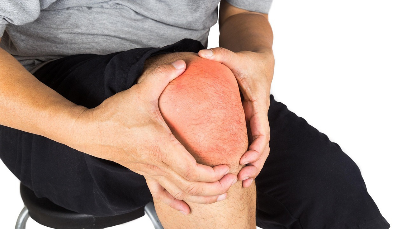
You’re performing a medial unicompartmental knee arthroplasty (mUKA). The medial compartment is bone-on-bone, the anterior cruciate ligament (ACL) is pristine, alignment is reasonable — and then you peek laterally.
There’s a little softening. Maybe some superficial fibrillation. An Outerbridge I or II lesion staring back at you from the lateral femoral condyle.
Do you abort? Convert to TKA? Or whisper reassuringly to yourself, “It’s fine…probably”?
According to one group of researchers, you can go ahead and relax. Mild lateral femoral condyle cartilage damage does not doom your medial UKA.
Let’s dive in.
The Central Clinical Question
Does mild cartilage damage (Outerbridge ≤ II) of the lateral femoral condyle negatively affect mid-term outcomes after medial UKA — or accelerate lateral compartment degeneration?
Lateral disease may be a contraindication, but what about mild disease? The kind that’s not full-thickness, not eburnated, not collapsing — just…imperfect.
This study tackled that exact gray zone.
A Lateral Compartment Reality Check
Between 2016 and 2020, 203 knees (177 patients) underwent medial UKA. Patients were divided into:
- Normal lateral cartilage
- Mild cartilage damage (Outerbridge I–II): Further subdivided by lesion location:
- Weight-bearing area
- Posterior weight-bearing area
- Medial side of the lateral condyle
Importantly, they excluded postoperative overcorrection of alignment and preoperative lateral meniscal extrusion.
So, no iatrogenic valgus overload and no dysfunctional lateral meniscus confounders.
Mean follow-up? A respectable 70.8 months (~6 years).
Function, Radiographic Progression and Survivorship
The research team collected data for: Hip-Knee-Ankle angle (HKA), Lateral compartment Kellgren-Lawrence (K-L) grade, Oxford Knee Score (OKS), Forgotten Joint Score (FJS), Kujala score, patient satisfaction and post-op complications.
The Headline Result: No Functional Penalty
Postoperatively the researchers found: OKS: No difference, FJS: No difference, Kujala score: No difference and patient satisfaction: Comparable across groups.
Bottom line: patients with mild lateral cartilage damage did just as well as those with pristine lateral compartments.
What About Radiographic Progression?
Of the 99 knees with mid-term standing radiographs 26.3% progressed one K-L grade and 73.7% remained unchanged.
But here’s the key: This mild radiographic progression did not translate into worse clinical outcomes.
And most importantly: No knees required conversion to TKA.
So, yes, some lateral compartments aged a bit radiographically — but they didn’t become clinically problematic.
Complications? Minimal.
Only 3 complications (1.5%), all in the normal cartilage group: 1 periprosthetic fracture, 1 bearing dislocation and 1 bearing rotation.
None required revision to TKA.
Notably, mild lateral cartilage damage was not associated with higher complication rates.
Why might this make biomechanical sense?
Medial UKA restores alignment without overcorrection (if done properly). If you avoid pushing patients into valgus, you’re not overloading the lateral side.
Add in intact ACL, functional lateral meniscus, no lateral meniscal extrusion, and no postoperative valgus overcorrection.
And that lateral compartment likely continues to share load physiologically.
Mild chondral changes may simply represent age-related cartilage wear rather than true compartmental disease.
The Real Take-Home: Surgical Judgment Still Matters
This study doesn’t mean “all lateral cartilage damage is fine.”
It specifically applies to Outerbridge I–II, no meniscal extrusion, no lateral joint space collapse, no inflammatory arthropathy and no valgus overcorrection.
The authors were careful. This wasn’t cavalier UKA indication creep — it was measured expansion into a very common gray area.
What This Means for Your OR Tomorrow
You’re in the joint. You see mild softening, some superficial fibrillation, no exposed bone, no lateral meniscal incompetence, and no fixed valgus.
Instead of converting to TKA out of fear, you can reasonably proceed with medial UKA — assuming alignment and meniscal function are appropriate.
That’s a big deal.
Because avoiding unnecessary TKA preserves bone stock, maintains native kinematics, shortens recovery and keeps future revision options open.
Limitations
This was a retrospective design, radiographs were available for only 99 of 203 knees, mean follow-up was 6 years (good, but not 15) and the population was homogenous (predominantly East Asian cohort).
Longer-term survivorship data will still be valuable. But mid-term? The data are reassuring.
Origin Study Title: Mild Lateral Femoral Condyle Cartilage Damage Does Not Affect the Outcomes of Medial Unicompartmental Knee Arthroplasty
Authors: Mincong Du, MSc; Zheng Li, MSc; Xufeng Jiao, MSc; Shuai An, M.D.; Xiaomei Yao, Ph.D.; Jiang Huang, M.D. Guobin Liu, M.D.; Guanglei Cao, M.D.; Ye Huang, M.D.