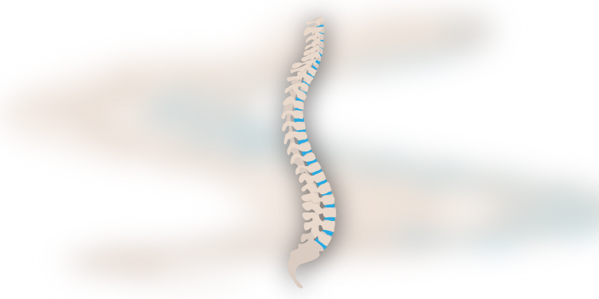
According to a new study from Washington University in St. Louis, Missouri and MD Anderson Cancer Center in Houston, Texas, preoperative diffusion basis spectrum imaging can predict improvement after surgical decompression for cervical spondylotic myelopathy with high accuracy for multiple outcome measures.
Preop Diffusion Basis Spectrum Imaging – How Effective?
2 min read Premium comments
#cervicalspondyloticmyelopathySecondary#diffusionbasespectrumimaging
The study, “Diffusion basis spectrum imaging predicts long-term clinical outcomes following surgery in cervical spondylotic myelopathy,” was published online on December 9, 2022 in The Spine Journal.
“A major shortcoming in improving care for cervical spondylotic myelopathy patients is the lack of robust quantitative imaging tools to guide surgical decision-making. Diffusion basis spectrum imaging, an advanced diffusion-weighted MRI technique, provides objective assessments of white matter tract integrity that may help prognosticate outcomes in patients undergoing surgery for cervical spondylotic myelopathy,” the researchers wrote.
For their study, the research group quantified the ability of diffusion basis spectrum imaging to predict important outcome measures at two-years follow-up in patients undergoing decompressive cervical surgery for cervical spondylotic myelopathy.
The team measured neurofunctional status outcomes using modified Japanese Orthopedic Association, MDI and DASH. They then measured patient quality-of-life using SF-36 PCS and SF-36 MCS. Finally, they also included the Neck Disability Index and North American Spine Society satisfaction index
The study group enrolled 50 patients and, using preoperative diffusion basis spectrum imaging metrics assessed white matter tract integrity through fractional anisotropy, fiber fraction, axial diffusivity, and radial diffusivity. They also used it to measure restricted and non-restricted to evaluate extra-axonal diffusion.
Finally, after enrolling their patients and collecting initial data, the team continued to follow-up for two years. They then used support vector machine classification algorithms to attempt to predict surgical outcomes at two-years follow-up.
In total, the study organizers enrolled 27 mild, 12 moderate and 11 severe cervical spondylotic myelopathy patients. Twenty-four patients underwent anterior decompressive surgery compared to 16 posterior approaches. In practice, the researchers were able to achieve 23.2 months mean (SD) follow-up (5.6, range 6.1-32.8).
After reviewing their data, the team concluded that using combined clinical and diffusion basis spectrum imaging data was most effective at predicting improvement.
In their final report, the researchers wrote, “These results suggest that diffusion basis spectrum imaging may serve as a non-invasive imaging biomarker for cervical spondylotic myelopathy valuable in guiding patient selection and informing preoperative counseling.”
Study authors include Justin K. Zhang, B.S., Dinal Jayasekera, Ph.D., Saad Javeed, M.D., Jacob K. Greenberg, M.D., Jacob Blum, B.S., Christopher F. Dibble, M.D., Ph.D., Sheng-Kwei Song, Ph.D. and Wilson Z. Ray, M.D., all of Washington University in Saint Louis, Missouri. Peng Sun, Ph.D. of UT MD Anderson Cancer Center, Houston, Texas also contributed to the study.
React:
Discussion
This is a fascinating development. In my practice we've seen similar outcomes with the revised protocol. The key differentiator seems to be patient selection criteria. Has anyone else noticed the correlation with BMI thresholds?
Great point. I'd push back slightly on the conclusion, the sample size in the cited study is too small to draw population-level inferences. That said, the directional signal is compelling and worth a larger RCT.
We implemented a similar approach last year. Early results are promising but we're still gathering 12-month follow-up data. Happy to share our protocol if anyone is interested.
Join the conversation
Orthopedic professionals are discussing this. Sign in and upgrade to read every comment and add your voice.