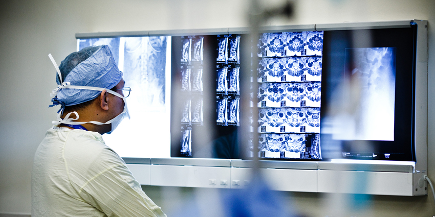
Putting preoperative, intraoperative, and postoperative radiographs side by side, researchers from the Rothman Orthopaedic Institute in Philadelphia asked the key question—can intraoperative lumbar lordosis and segmental lordosis predict postoperative improvement?
How Well Can Segmental Lordosis Predict Outcomes?
2 min read Premium comments
Secondary#anteriorlumbarinterbodyfusion#lumbarlordosis#lumbarsagitalalignment
Their study, “A Short-Term Assessment of Lumbar Sagittal Alignment Parameters in Patients Undergoing Anterior Lumbar Interbody Fusion,” appears in the December 1, 2022, edition of Spine.
“The main reason we looked at this topic,” stated co-author Mark Lambrechts, M.D., “was to figure out if there was any real value in intraoperative radiographs aside from identifying if the instrumentation and grafts appear appropriately placed.”
“We assumed lumbar lordosis and segmental lordosis after the anterior lumbar interbody fusion (ALIF) would be similar between the post-instrumentation intraoperative X-rays and the postoperative radiographs, but we felt we needed evidence to prove this.”
Mining the electronic medical records of Thomas Jefferson University Hospital, the researchers looked at patients over the age of 18 who had single- and two-level anterior lumbar interbody fusion with posterior instrumentation between 2016 and 2020. Of the 118 patients who met the inclusion criteria, 75 had one-level fusions and 43 had a two-level fusion.
The team determined that lumbar lordosis significantly increased following on-table positioning.
In addition, lumbar lordosis significantly decreased between the intraoperative to postoperative radiographs at two to six weeks, while no change was identified between the intraoperative and more than three-month postoperative radiographs.
The Rothman group found that sagittal lordosis significantly increased from the preoperative to intraoperative radiographs, but later decreased at the two to six weeks follow up and at the final follow up.
“The most interesting result,” Dr. Lambrechts told OTW, “is that the post-instrumentation intraoperative radiographs are more accurate predictors of lumbar lordosis and segmental lordosis than the immediate postoperative images.”
“This is possibly due to a couple of reasons including postoperative pain/splinting or subconscious posturing in patients due to neural element compression which helps relieve pain in the preoperative period (which still probably persists in the early postop period).”
“One of the most interesting findings is that at around 1-year follow up the segmental lordosis correction achieved with the ALIF diminishes but the amount of segmental lordosis lost was relatively predictable when calculating the amount of allograft resorption or cage subsidence since each mm or resorption/subsidence led to approximately 0.5 degrees loss of segmental lordosis.”
“Although this manuscript does shed some light on the value of intraoperative imaging, we still have a poor understanding of what appropriate segmental/lumbar lordosis correction is needed in individual patients. Each patient has a specific lumbar distribution index, which is difficult to determine after significant disc degeneration has occurred. Over or under-correction of a patient’s lordosis may alter a patient’s postoperative outcome, but we still do not have a great understanding of this topic.”
React:
Discussion
This is a fascinating development. In my practice we've seen similar outcomes with the revised protocol. The key differentiator seems to be patient selection criteria. Has anyone else noticed the correlation with BMI thresholds?
Great point. I'd push back slightly on the conclusion, the sample size in the cited study is too small to draw population-level inferences. That said, the directional signal is compelling and worth a larger RCT.
We implemented a similar approach last year. Early results are promising but we're still gathering 12-month follow-up data. Happy to share our protocol if anyone is interested.
Join the conversation
Orthopedic professionals are discussing this. Sign in and upgrade to read every comment and add your voice.