Could a magnetic field and hydrogels arrest cartilage degeneration, if not also regenerate the tissue? That’s what a team of researchers in the Perelman School of Medicine at the University of Pennsylvania have just studied and their conclusions are very encouraging. Their study, “Magneto‐Driven Gradients of Diamagnetic Objects for Engineering Complex Tissues,” appears in the October 19, 2020 edition of Advanced Materials.
Stunning New Cartilage Repair Research From U Penn
2 min read Premium comments
Secondary#cartilagerepair#perelmanschoolofmedicine
“We found that we were able to arrange objects, such as cells, in ways that could generate new, complex tissues without having to alter the cells themselves,” said the study’s first author, Hannah Zlotnick, a graduate student in bioengineering who works in the McKay Orthopaedic Research Laboratory at Penn Medicine.
“Others have had to add magnetic particles to the cells so that they respond to a magnetic field, but that approach can have unwanted long-term effects on cell health. Instead, we manipulated the magnetic character of the environment surrounding the cells, allowing us to arrange the objects with magnets.”
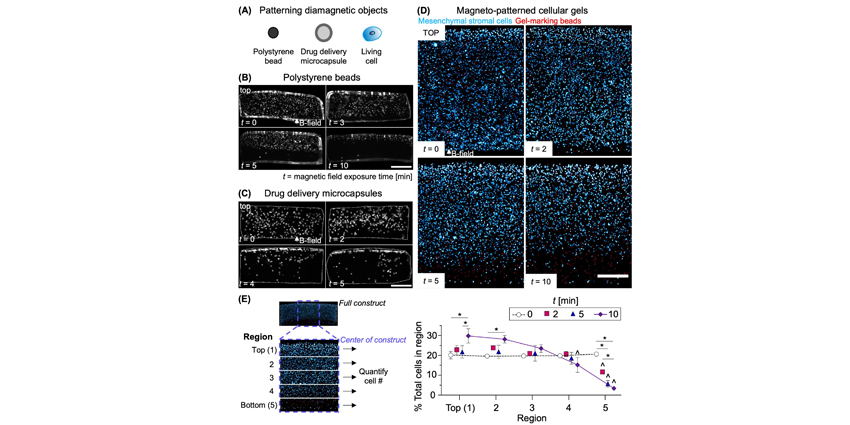
The researchers, including Robert Mauck, Ph.D., director of the McKay Lab, added a magnetic liquid to a 3D hydrogel solution. The result was that cells and other non-magnetic objects—including drug delivery microcapsules—were able to be arranged into patterns that mimicked natural tissue through the use of an external magnetic field.
The authors wrote, “After brief contact with the magnetic field, the hydrogel solution (and the objects in it) was exposed to ultraviolet light in a process called ‘photo crosslinking’ to lock everything in place, and the magnetic solution subsequently was diffused out. After this, the engineered tissues maintained the necessary cellular gradient.”
Zlotnick told OTW, “To date, there is no clinical cartilage repair technique that has been shown to recapitulate the unique biologic structure of the tissue. We believe that our magneto-patterning strategy is particularly exciting, because it allows us to engineer tissues with cellular gradients, similar to native tissues. Therefore, in the lab, we can create cartilage tissue replacements that better resemble native cartilage, and may therefore better integrate in vivo.”
Regarding their next steps, Zlotnick said, “We are currently planning an upcoming large animal study to implant these engineered cartilage tissues in cartilage defects of living animals. It is essential to assess the ability of our magneto-patterned tissues to withstand joint-scale forces. We are excited to translate this technology to the next stage of testing.”
React:
Discussion
This is a fascinating development. In my practice we've seen similar outcomes with the revised protocol. The key differentiator seems to be patient selection criteria. Has anyone else noticed the correlation with BMI thresholds?
Great point. I'd push back slightly on the conclusion, the sample size in the cited study is too small to draw population-level inferences. That said, the directional signal is compelling and worth a larger RCT.
We implemented a similar approach last year. Early results are promising but we're still gathering 12-month follow-up data. Happy to share our protocol if anyone is interested.
Join the conversation
Orthopedic professionals are discussing this. Sign in and upgrade to read every comment and add your voice.