For the first time, a study has shown visual evidence of biofilm during “aseptic” revision spine surgery. The article, “High Prevalence of Biofilms on Retrieved Implants from Aseptic Pseudarthrosis Cases,” was published by the Japanese Society for Spine Surgery and Related Research. Aakash Agarwal, Ph.D., and colleagues Megan Mooney, Ashish G. Agarwal, Daksh Jayaswal, Gayane Saakyan, Vijay Goel, Jeffrey C. Wang, Neel Anand, Steve Garfin, Vithal Shendge and Hossein Elgafy authored the study.
Landmark Study: 1st Visual Biofilm Evidence From “Aseptic” Surgery
5 min read Premium comments
#asepticrevisionsurgery#implantbiofilm#occultinfection
Researchers Find Visual Evidence of Biofilm
What inspired the researchers to conduct this study?
Aakash Agarwal, Ph.D., who is Director of Research at Spinal Balance AND a leading researcher in the Departments of Bioengineering and Orthopaedic Surgery at the University of Toledo, had observed that recent research has shown cultures of bacteria and high correlation between presence of bacteria during supposedly “aseptic revision surgery.” Two such studies, published in European Spine Journal and Journal of Neurosurgery, also showed strong evidence of correlation between bacteria presence and screw loosening.
Agarwal and his colleagues decided to follow up on this research, as well as their own previous research, which indicated that orthopedic surgeons are impregnating bacterial biodose in patients through use of repeatedly reprocessing and intraoperatively exposing implants during surgery. The subsequent research results showed, for the very first time, documented evidence of surreptitious biofilm in patients requiring “aseptic” revision surgery (with no clinical signs of infection).
Agarwal said, “For the past couple of years, numerous leading researchers around the world have shown evidence regarding higher prevalence of positive microbial cultures in revision spine surgeries… Many of these researchers (especially the recent ones) also reported higher correlation of these positive microbial cultures with pseudarthrosis, or screw loosening. These past authors have used the terms: occult infection, chronic implant infection etc. to describe this scientific observation.”
Catalysts for the Research
This caused Agarwal and his colleagues to wonder why this was happening and what the root cause of such infection might be. He stated, “the answer to which I have pointed out in my previous collaborative work, [is] that most implants are undergoing contamination both preoperatively and intraoperatively. The solution to this is two-step asepsis. Unless we adhere to two-step asepsis and avoid these known modes of contamination during spine surgery, there is no scientific way to identify additional sources of risk factors that may be associated with it.”
A second concern of the researchers was the serious risks of such secondary infections.
Agarwal explained that much previous research has already associated biofilm infection after revision surgery with late onset infection and/or pseudoarthrosis. The current body of research on this topic, according to Agarwal, is “excellent, and the only reason the scientific community have not completely digested the enormous body of work is because they worry that it is easy to contaminate retrieved study samples and have false positives.”
“However, if you can demonstrate presence of biofilm on implants from failed spine surgery via visual means, it strongly supports the studies previously conducted by all these researchers showing positive culture.”
Biofilm Defined
What exactly is biofilm? How does it function?
Agarwal told OTW, “Biofilm is an aggregate of microorganisms in which cells that are embedded within a self-produced matrix of extracellular polymeric substances, adhering to each other and onto the implant surface. It is a system that helps bacteria survive in unfriendly conditions (like our body) by making its own suitable environmental conditions upon a host (implants).”
“Biofilm is a real problem and a common cause of implant failure, with serious consequences. The chronically infected implants cannot be treated without removal of the implant, as the biofilm protects the bacteria against antibiotics,” Agarwal said.
This new, visual evidence of biofilm on implants from patients undergoing “aseptic” revision surgery, as opposed to in vitro or animal models, makes the reality of biofilm more concrete.
The Study
The researchers noted in their work that, “Recent literature showing pseudarthrosis and pedicle screw loosening with subchronic infection at the pedicle of the vertebra,” and that “The positive culture results of a previous retrieval analysis show that such patients have a high frequency of bacterial contamination.” The researchers described the objective of the study as ”to visually capture the architecture of these undiagnosed infections.”
Revision surgery is common after spinal fusion procedure. Known causes include surgical site infection (SSI), implant-related failure, pseudarthrosis, degenerative disease progression, adjacent segment diseases, nerve impingement recurrence, incorrect initial diagnoses and surgical error.
The researchers cite risk factors for recurrent back pain as smoking, obesity, smoking, autoimmune disease, and diabetes, all of which can inhibit spinal fusion.
Aseptic screw loosening is common. The researchers noted in their study that although “it was previously deemed a mechanical failure at the screw–bone interface in the absence of solid fusion, recent studies have highlighted a possible biological pathway.” Orthopedic researchers have theorized a biological path, with bacterial growth surrounding implant but no known clinical symptoms. As the researchers noted, “This is consistent with other studies that demonstrated a positive bacterial biodose impregnation at the screw–bone interface during spine surgery.”
Previous studies have pointed toward high rates of late-onset infection after instrumented spine surgery, which is consistent with the researchers’ hypothesis of biological pathway as a cause. However, the researchers wrote, “the exact architecture of such implant associated biofilms is yet to be determined because all previous studies have relied on bacterial cultures, wet-lab procedures, or animal studies.”
The researchers aimed to characterize what is assumed to be “aseptic” pedicle screw loosening in revision surgery patients and to “determine both the frequency and visual architecture of biofilms on implant surfaces.”
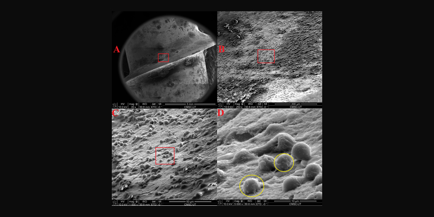
The pedicle screw explants from 10 individual patients who had undergone spine revision surgery for pseudoarthrosis were imaged through scanning electron microscopy and X-ray spectroscopy. The detailed architecture of biofilm on these was then evaluated. Eight patient swabs from implant tissues were also sent for cultures to assess for bacterial infiltration in the tissues beyond any biofilm. Samples with current or previous clinically diagnosed infection and/or mechanical failure of the implant due to physical trauma were excluded.
The results were that the study was able to capture images of the visual architecture of the biofilm on implants, with 77% of pseudarthrosis cases presented with loose pedicle screws. These cases were diagnosed by preoperative computed tomography scan. The scan showed radiolucency along the screw track. These were subsequently confirmed intraoperatively, with 72% of the cases showing explant biofilm.
The researchers concluded that even without clinical indication of infection, “impregnated bacteria could form a biofilm around an implant, and this biofilm can remain undetected via contemporary diagnostic methods, including swabbing…Implant biofilm is frequently present in ‘aseptic’ pseudarthrosis cases.”
Research Implications
“A previous hypothesis suggests that in several susceptible patients, the bacteria from initial surgery could lie dormant and thrive via biofilm formation on the implant. The current study provides preliminary data that support this hypothesis as a possible onset mechanism for delayed and late infection,” the researchers wrote.
Agarwal and his research team look forward to possible positive implications for patient outcomes. Agarwal said, “Finding a cure for biofilm will involve a lot of cost and time. Therefore, first and foremost, we should immediately start using the preventative measures to avoid the implants from unnecessary contamination. We have been underestimating the power of prevention. Further future research should focus on establishing high efficacy and safety for new biomaterials, coatings, and innovative biofilm-disrupting-treatments such as electromagnetic or other non-invasive methods.”
The researchers also noted, “This study also highlights the importance of keeping screw/screw–bone interfaces devoid of bioload because of the propensity of bacterial inoculation to form biofilms around the implant. Such biofilms can remain undetected by contemporary diagnostic methods, including swabbing.”
React:
Discussion
This is a fascinating development. In my practice we've seen similar outcomes with the revised protocol. The key differentiator seems to be patient selection criteria. Has anyone else noticed the correlation with BMI thresholds?
Great point. I'd push back slightly on the conclusion, the sample size in the cited study is too small to draw population-level inferences. That said, the directional signal is compelling and worth a larger RCT.
We implemented a similar approach last year. Early results are promising but we're still gathering 12-month follow-up data. Happy to share our protocol if anyone is interested.
Join the conversation
Orthopedic professionals are discussing this. Sign in and upgrade to read every comment and add your voice.