We recently covered the first use of Augmedics Ltd ’s xvision augmented reality system for spine surgery as well as the company’s recent fundraising success.
Rush University Amazed at Revolutionary MIS Vision Tool
2 min read Premium comments
#minimallyinvasivespinesurgery#augmedics#xvisionspinesystem
Since publication of our last story, OTW had a chance to interview Frank Phillips, M.D. Phillips, a professor and director of the Division of Spine Surgery and the Section of Minimally Invasive Spine Surgery at Rush University Medical Center in Chicago, Illinois, who used xvision on June 15, 2020 to assist in placing pedicle screws for a minimally invasive L3-S1 anterior/posterior lumbar fusion revision.
When asked about his initial thoughts of using the xvision system, Phillips simply said, “Amazing.” When probed further, he went on to describe the “game changing technology.” Phillips was part of the preclinical study using cadavers, so the system was not unfamiliar. The results of that study were used to support the FDA 510(k) clearance granted to the system in December 2019.
Phillips acknowledged that the benefits of the system over traditional navigation are not earthshattering for open procedures where the surgeon can see the portion of the spine that he or she is working on. The main benefit for open procedures is that the instrument and implant visualization is overlaid on the surgeon’s headset display, eliminating the need to look up at a monitor while placing screws or implants.
However, Phillips explained that the system really excels when it comes to minimally invasive procedures.
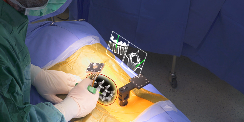
Like current navigation systems preoperative CT, or MRI are fed into the system and instruments are registered in a similar fashion. The xvision provides visualization of the instruments and implants directly over the preoperative imaging both on the surgeons’ headsets, as well as on a computer screen or monitor in the OR.
Where the xvision differs from current navigation systems is the 3D projection of the spine on the surgeons’ headsets. Without looking up a surgeon can see the patient’s spine in 3 dimensions and the instruments and implants being used right where the patient’s spine really exists in space. Another benefit to being able to see the skin and anatomy underneath, Phillips reports having better skin incisions, with less maneuvering since you can visualize the entire surgical corridor before you start cutting.
Phillips said the system is “very intuitive” because you are looking right at the patient, you just happen to see everything through the skin as if you were performing an open procedure. When two surgeons are performing a procedure, each will see instruments being used and the spine from his or her own perspective. The OR monitor does not display in 3D but allows OR staff to rotate the image to change their perspective of the surgical site.
While the company does not make any claims regarding the speed of placing pedicle screws with the system, Phillips told OTW that he was able to place 4 screws in 12 minutes reducing his overall OR time, compared with other navigation systems. The company claims a 98.9% accuracy rate when placing pedicle screws based on the cadaver study published in 2019.
When asked what his future plans are regarding use of the xvision system, Phillips replied that he plans to use it in most of his upcoming minimally invasive surgeries.
React:
Discussion
This is a fascinating development. In my practice we've seen similar outcomes with the revised protocol. The key differentiator seems to be patient selection criteria. Has anyone else noticed the correlation with BMI thresholds?
Great point. I'd push back slightly on the conclusion, the sample size in the cited study is too small to draw population-level inferences. That said, the directional signal is compelling and worth a larger RCT.
We implemented a similar approach last year. Early results are promising but we're still gathering 12-month follow-up data. Happy to share our protocol if anyone is interested.
Join the conversation
Orthopedic professionals are discussing this. Sign in and upgrade to read every comment and add your voice.