If a surgeon can “see” the problem, he or she is likely to fix it.
FDA-Cleared Carestream Radiography System Promises Enhanced Clarity
2 min read Premium comments
#fda510kclearanceSecondary#xrays#carestream#radiography
Carestream Health, Inc.’s recently FDA 510(k)-cleared DRX-Evolution Plus with Carestream Digital Tomosynthesis (DT) promises more clarity, simplified workflow and reduced exam time from standard X-rays, according to a January 14, 2020 company press release.
The DT system is a digital radiography system that performs a wide range of general radiographic exams.
The FDA clearance document states “The device is a permanently installed diagnostic x-ray system for general radiographic x-ray imaging included tomography. This device also supports digital tomosynthesis…” but is “…not to be used for imaging pediatric patients.”
Tomosynthesis Clarity
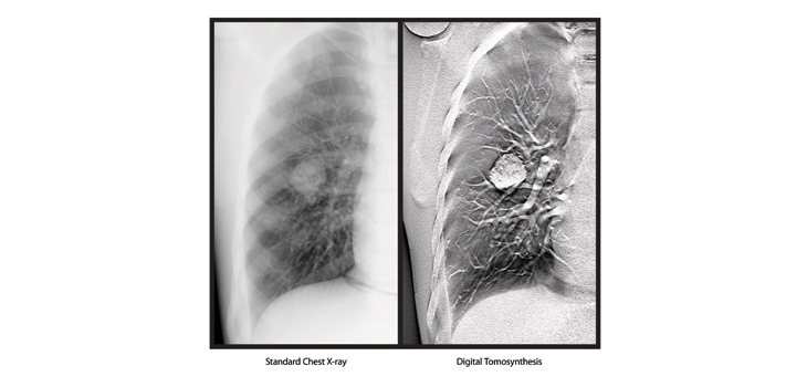
“Tomosynthesis is an advanced radiographic application that produces individual coronal “slice” images through an anatomical region of interest (ROI). The FDA documents explains that the slices are produced through multiple projection radiographic images that are acquired in rapid succession as the X-ray tube sweeps and rotates across the ROI. Once acquired these projection images are subject to processing that registers and reconstructs them to individual tomographic slices.”
As a result, claims the company, the system can generate data from a series of low dose X-ray images of the same organ, taken at the same X-ray exposure, from different angles.
The clarity is achieved “By establishing the initial pivot level (fulcral plane) and exposing across the sweep angle the anatomical details in the fulcral plane are persevered, while anatomical clutter above or below the plane will be blurred. Both linear tomography and digital tomosynthesis isolate the anatomy of interest in the fulcral plane from unwanted clutter and improve the contrast to noise ratio of the image details.”
The FDA document notes the sweep imaging technique “generates multiple two-dimensional (2D) coronal slices (i.e. planes) from a series of low dose x-ray images of the same anatomy taken at the same exposure but at different angles. During a tomosynthesis acquisition the detector will be stationary while the tube head travels (sweeps) in a straight path (i.e. focal spot travel path). For each exposure, the tube will be angled toward the center of the detector.”
Sarah Verna, Worldwide Marketing Manager for Global X-ray Solutions at Carestream, said doctors and radiologists will be able to “perform scans quickly, providing valuable clinical information for further diagnosis while improving the workflow in an X-ray room.”
“In trauma centers, CT rooms are always overbooked and there’s always a wait,” Verna continued. “Now a doctor can further examine any body part.”
React:
Discussion
This is a fascinating development. In my practice we've seen similar outcomes with the revised protocol. The key differentiator seems to be patient selection criteria. Has anyone else noticed the correlation with BMI thresholds?
Great point. I'd push back slightly on the conclusion, the sample size in the cited study is too small to draw population-level inferences. That said, the directional signal is compelling and worth a larger RCT.
We implemented a similar approach last year. Early results are promising but we're still gathering 12-month follow-up data. Happy to share our protocol if anyone is interested.
Join the conversation
Orthopedic professionals are discussing this. Sign in and upgrade to read every comment and add your voice.