mediCAD Hectec GmbH, based near Landshut, Germany, has announced that its newest version of mediCAD Spine 3D now includes full support for MRI scans.
mediCAD Announces Upgrade to mediCAD Spine 3D
2 min read Premium comments
Secondary#digitalpreoperativeplanning#medicadspine3d
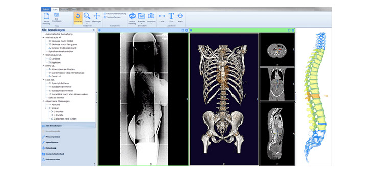
According to the company, “Previously, physicians could use mediCADSpine 3D to plan with x-rays and CT scans. In this new release of mediCAD Spine 3D, Version 2.2, the software now offers full MRI support with complete recognition of all bony structures. The new version also adds Le Huec sagittal balance functionality and an even better and faster automatic segmentation feature.”
“Of course, as with previous versions, mediCADSpine 3D includes a constantly growing database of digitized templates obtained directly from the world’s leading implant manufacturers…Across mediCAD Hectec GmbH’s entire product line, physicians are able to readily access around 3,000 implants from 130 international implant manufacturers.”
Patryk Ploschka, product manager of mediCAD Spine 3D, told OTW, “The high field strength of the MRI enables the precise diagnosis of even small structures. With normal X-ray images, the condition of the spine cannot really be determined: one recognizes the cartilage thickness only indirectly.”
“An MRI scan can diagnose diseases of the spine. Depending on the extent of the back pain, the cervical, thoracic or lumbar spine optimizes high-resolution and thin-layer images in top quality by optimizing the respective examination.”
“On the basis of these exact images a reliable diagnosis can be made, and an ideal and individual treatment can be initiated. In addition to the representation of the bones, muscles and ligaments of the entire spine, if necessary, the spinal cord can also be examined in the entire course (from the cervical cord to the transition of the spinal cord into the individual nerve fibers). MRI opens up the possibility of non-invasively diagnosing the condition of the spine (or any other part of the body) with maximum precision without exposure to radiation. This results not only in diagnostic findings, but also consequences for the treatment.”
“MRI scans are standard within spine-surgery as MRI can show soft tissues and spinal cord in great detail with a very high contrast between the different tissues. In this way, within a non-invasive radiation-free examination, MRI can visualize all structures of the spine and intervertebral discs, the parts of the nervous system lying there and the surrounding soft tissues with high quality.”
“Constrictions of the spinal canal or the nerve exit points from the spine are reliably detected. Therefore, with mediCAD Spine 3D now being able to work with MRI scans (as well as with X-ray and CT), mediCAD Hectec GmbH once again shows why it is one of the world market leaders in the field of digital preoperative planning in surgical orthopedics.”
React:
Discussion
This is a fascinating development. In my practice we've seen similar outcomes with the revised protocol. The key differentiator seems to be patient selection criteria. Has anyone else noticed the correlation with BMI thresholds?
Great point. I'd push back slightly on the conclusion, the sample size in the cited study is too small to draw population-level inferences. That said, the directional signal is compelling and worth a larger RCT.
We implemented a similar approach last year. Early results are promising but we're still gathering 12-month follow-up data. Happy to share our protocol if anyone is interested.
Join the conversation
Orthopedic professionals are discussing this. Sign in and upgrade to read every comment and add your voice.