New multicenter research has found that a detailed preoperative spine classification has improved outcome predictions for patients with adolescent idiopathic scoliosis (AIS).
Scoliosis Classification System Teases Out Patient Differences
2 min read Premium comments
Secondary#spinaldeformity#adolescentidiopathidscoliosis#sabapasha
The work, “A hierarchical classification of adolescent idiopathic scoliosis: Identifying the distinguishing features in 3D spinal deformities,” was published in the March 20, 2019 edition of PLoS One.
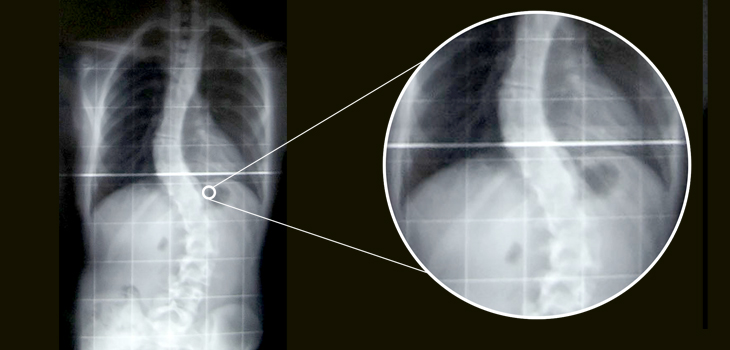
Co-author Saba Pasha, Ph.D. told OTW, “A subtype of pediatric scoliosis, known as adolescent idiopathic scoliosis, is a three-dimensional spinal deformity. Because the spinal X-rays have been used conventionally to monitor these patients, the guidelines for surgical planning uses 2-dimentional information that can be measured on the X-rays.”
“This has resulted in poor surgical outcomes because the detailed differences between the spines cannot be captured via 2D X-ray measurements. Our method used 3D models of the spine to develop a new classification but then provides a list of detailed differences that can be captured from the 2D X-rays. This helps the physician to look for those parameters on the X-rays to better identify the differences between the patients.”
The authors wrote, “A total number of 103 right thoracic left lumbar pre-operative AIS patients were included retrospectively and consecutively. A total number of 20 non-scoliotic adolescents were included as the control group. All patients had biplanar X-rays and 3D reconstructions of the spine…”
Dr. Pasha told OTW, “We found five subgroups of patients who are classified under one group using the current clinically used classification. Our finding shows that considering the alignment of the spine in 3D is necessary because otherwise many subtle details that differentiate one patient from another may be ignored. The new classification allows for the determination of patient-specific treatment which in turn can improve the outcomes of surgical intervention. More specifically, our results found 5 subgroups of scoliosis with a similar 2D curve but significantly different 3D spinal curves, which shows the level of details that can be extracted using our classification.”
“As spinal surgery in scoliotic patients aims to correct the deformity and ‘normalize’ the spine, a better understanding of the deviation of the spine from the normative spine curvature can inform the surgeon about the changes that are needed to be imparted during the surgery. Our classification allows the surgeon to acknowledge this difference and plan better for surgery. Thus far, this was only possible through a complicated and time-consuming image processing, but our method allows us to obtain the same information from the clinically available X-ray images. Learning about this new classification led the surgeon to incorporate that very sought-after third dimension of the spine, which is traditionally hard to access in clinic, in their clinical practice.”
React:
Discussion
This is a fascinating development. In my practice we've seen similar outcomes with the revised protocol. The key differentiator seems to be patient selection criteria. Has anyone else noticed the correlation with BMI thresholds?
Great point. I'd push back slightly on the conclusion, the sample size in the cited study is too small to draw population-level inferences. That said, the directional signal is compelling and worth a larger RCT.
We implemented a similar approach last year. Early results are promising but we're still gathering 12-month follow-up data. Happy to share our protocol if anyone is interested.
Join the conversation
Orthopedic professionals are discussing this. Sign in and upgrade to read every comment and add your voice.