A new study, “Longitudinal Strain Measures of White Matter Tracts in Youth Football Players,” based on MRI scans presented at the recent annual meeting of the Radiological Society of North America suggests that repetitive blows to the head result in brain changes among youth football players.
Youth Football Changes Nerve Fibers in Brain
2 min read Premium comments
Secondary#concussion#subconcussivesymptoms#youthfootball
While most football teams have concussion protocols to remove players who show signs of concussion from the game, subconcussive symptoms are harder to determine. Because many hits to the head are subconcussive (meaning they are below the threshold of a concussion), there is a driving concern to determine the long-term consequences of subconcussive hits to the head.
“The years from age 9 to 12 are very important when it comes to brain development,” said lead author Jeongchul Kim, Ph.D., of Wake Forest School of Medicine in Winston-Salem, North Carolina.
“The functional regions of the brain are starting to integrate with one another, and players exposed to repetitive brain injuries, even if the amount of impact is small, could be at risk.”
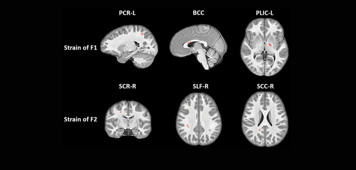
To determine the effects of these subconcussive collisions on youth football players, Kim and his colleagues used a novel MRI method that looks at the strain evident on white matter tracts, the bundles of nerve fibers that carry information between different areas of the brain.
“The focus here was on deformations of these fiber bundles,” Kim said. “Changes from collisions might cause elongation or contraction of these bundles.”
The MRI studies were done on 26 male youth football players, the average age being 12 years old. The studies were performed before the season had begun and again about three months after the end of the season. Their results were compared to 22 boys of similar age who did not participate in contact sports.
According to the data collected, the football players developed changes in the corpus callosum, an important band of nerve fibers that connects the two halves of the brain. It is responsible for integrating cognitive, motor and sensory functions between the two sides of the brain.
Signs of greater axial strain (contraction) were observed in some parts of the corpus callosum while indications of radical strain (expansion) were seen in other parts.
“The body of the corpus callosum is a unique structure that’s somewhat like a bridge connecting the left and right hemispheres of the brain,” Kim added.
“When it’s subjected to external forces, some areas will contract and others will expand, just like when a bridge is twisting in the wind.”
Kim explained that their findings suggest that repetitive head impacts associated with participation in youth contact sports could lead to changes in the shape of the corpus callosum during this critical time of brain development, however more evidence is needed to confirm their findings.
He and his colleagues will continue to study the players to see if any additional deformations occur with the ultimate goal of providing guidelines for safe football play.
“It’s best to detect changes at the earliest possible time,” Kim said.
The Radiological Society of North America’s 2018 annual meeting was held between November 20 and November 30 in Chicago.
React:
Discussion
This is a fascinating development. In my practice we've seen similar outcomes with the revised protocol. The key differentiator seems to be patient selection criteria. Has anyone else noticed the correlation with BMI thresholds?
Great point. I'd push back slightly on the conclusion, the sample size in the cited study is too small to draw population-level inferences. That said, the directional signal is compelling and worth a larger RCT.
We implemented a similar approach last year. Early results are promising but we're still gathering 12-month follow-up data. Happy to share our protocol if anyone is interested.
Join the conversation
Orthopedic professionals are discussing this. Sign in and upgrade to read every comment and add your voice.