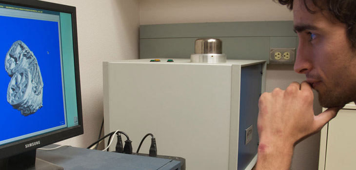
John A. Szivek, Ph.D., a biomedical engineer and professor of orthopedic surgery at the University of Arizona (UA), has received a five-year, $2 million grant from the U.S. Department of Defense (DoD) to learn how to heal bone fractures using a combination of 3D printing and adult stem cells.
$2 Million DoD Grant for 3D Printing and Stem Cells
2 min read Premium comments
Secondary#stemcells#3dprintingbigbonedefects#departmentofdefensegrant
“Imagine an impact that causes half of a long bone to shatter so that it can’t be put back together. No current surgical treatment,” he explained, “can ensure that kind of injury will heal. Injuries can cause big bone defects.”.
The unfortunate fact that Szivek is confronting is that not all broken bones will heal. At the University of Arizona he hopes to solve that problem using a combination of 3D printing and adult stem cells.
Szivek’s plan is to 3D print plastic bone-shaped frames that can fit into the voids left by missing or broken bone segments. He then plans to fill the scaffolds with calcium particles and adult stem cells.
“Once it has been implanted, the scaffold will serve as a template for the bone to grow on”, Szivek explained.
Szivek ran some pilot studies which showed the DoD that this technique works well. “We achieved complete bone formation, covering a large bone defect in about three months. Now we want to make that healing process even faster,” he said. “The team will test whether exercise early in the healing process can help speed up healing and recovery.”
“Studies have shown that exercise makes your bones grow, so maybe we can make bone on our scaffolds grow even faster with exercise,” Szivek said.
“To test this theory, 3D implants will be embedded with tiny sensors that can wirelessly transmit exercise activity. These sensors will analyze loading, or how much weight is being put on the scaffold, and for what length of time.”
“Bone size changes in a regularly exercising group will be compared to an inactive group. Szivek’s team have hypothesized that the actively exercising group will show more rapid bone growth. His team also hopes to develop guidelines for post-surgical physical therapy by demonstrating that exercise leads to better bone formation.”
“Patients often re-break the damaged bone area after surgeons try to repair it and the limb will eventually be amputated,” Szivek explained.
“If his study succeeds, Szivek hopes to expand his testing with clinical trials which would enroll military personnel.”
UA President Robert C. Robbins, M.D., commended Szivek’s lab: “This is an incredible example of the kind of innovative research that is made possible by technological advancement through the convergence of the biological, physical and digital worlds, and exactly the kind of project that demonstrates how the UA is a leader in cutting-edge solutions to difficult challenges.”
“The work that Dr. Szivek and his team are doing to help these individuals is a great example of using new technology to significantly improve quality of life for patients. I am confident their unique research will lead to the development of more effective treatments to repair critical bone injuries.”.
Szivek also hopes the potential therapy also will help patients with bone cancers that have been removed operatively.
React:
Discussion
This is a fascinating development. In my practice we've seen similar outcomes with the revised protocol. The key differentiator seems to be patient selection criteria. Has anyone else noticed the correlation with BMI thresholds?
Great point. I'd push back slightly on the conclusion, the sample size in the cited study is too small to draw population-level inferences. That said, the directional signal is compelling and worth a larger RCT.
We implemented a similar approach last year. Early results are promising but we're still gathering 12-month follow-up data. Happy to share our protocol if anyone is interested.
Join the conversation
Orthopedic professionals are discussing this. Sign in and upgrade to read every comment and add your voice.