A new retrospective analysis sought to determine the most effective way of diagnosing unilateral sacroiliitis, a rare inflammatory disease.
Unilateral Sacroiliitis: MRI or Tissue Studies for Diagnosis?
2 min read Premium comments
Secondary#subchondralmarrowedema#unilateralsacrilitis
The study, “Unilateral sacroiliitis: differentiating infective and inflammatory etiology by magnetic resonance imaging and tissue studies,” appears in the October 23, 2018 edition of the European Spine Journal.
Co-author Rishi Mugesh Kanna, M.S. (Orthopaedics), M.R.C.S. (Ed), F.N.B. Spine Surgery with the Department of Orthopaedics, Traumatology and Spine Surgery, Ganga Hospital in Coimbatore, India, explained the issues and need for his study to OTW, “Developing countries like India have a high incidence of infectious diseases. While most infectious diseases have a typical pattern of presentation and fairly defined methods of evaluation and treatment, spinal infectious diseases can be an enigma.”
“Its clinical presentation may mimic non-infectious inflammatory and degenerative conditions where the management is diametrically opposite. Hence, there is an increasing reliance on imaging studies to diagnose infectious diseases with more dependability and clarity.”
“Unilateral sacroiliitis, though uncommon, has a varied etiology, and MRI plays an important role in the diagnosis. The main aim of the study was to evaluate the efficacy of MRI parameters in differentiating infective and inflammatory causes of unilateral sacroiliitis.”
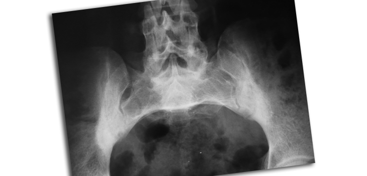
The authors described their methodology as follows: “In total, 33 patients (mean age—33.4 ± 17.2 years) with MRI features of unilateral sacroiliitis had presented with unilateral gluteal pain (100%) and positive Patrick’s test (91.9%).”
“Based on the MRI features of severe subchondral marrow edema, widening of joint space, intra-articular abscess and periarticular muscle abscess, infective sacroiliitis was diagnosed in 20/33 (60.6% cases). A total of 13/33 (39.3%) patients had features of inflammation based on the following MRI criteria:
- subchondral sclerosis with minimal edema
- erosions
- maintained joint space without abscess/destruction.”
Dr. Kanna added, “We observed that MRI had good sensitivity (71%) and 100% specificity in diagnosing inflammatory sacroiliitis. It had low specificity, but 100% sensitivity for diagnosing infective sacroiliitis.”
“But specific MRI findings could not differentiate pyogenic and tubercular etiology in patients with infective sacroiliitis. Hence, we concluded that patients diagnosed as inflammatory sacroiliitis on MRI are unlikely to benefit from further tissue studies, while biopsy is recommended in patients diagnosed on MRI as infective sacroiliitis to confirm the diagnosis and also to isolate the organism through tissue studies.”
“MRI features including sub-chondral sclerosis with minimal edema, erosions, maintained joint space without abscess/destruction were the diagnostic criteria for inflammatory sacroiliitis which had 100% specificity.”
“Based on the MRI findings, patients diagnosed with inflammatory sacroiliitis on MRI may not require tissue studies and can possibly be treated as having inflammatory arthritis. But a percutaneous biopsy is recommended in patients diagnosed by MRI as having infective sacroiliitis to initiate anti-microbial therapy based on the isolated organism.”
React:
Discussion
This is a fascinating development. In my practice we've seen similar outcomes with the revised protocol. The key differentiator seems to be patient selection criteria. Has anyone else noticed the correlation with BMI thresholds?
Great point. I'd push back slightly on the conclusion, the sample size in the cited study is too small to draw population-level inferences. That said, the directional signal is compelling and worth a larger RCT.
We implemented a similar approach last year. Early results are promising but we're still gathering 12-month follow-up data. Happy to share our protocol if anyone is interested.
Join the conversation
Orthopedic professionals are discussing this. Sign in and upgrade to read every comment and add your voice.