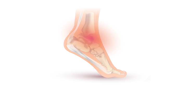
Mount Sinai researchers recently discovered a new, minimally invasive technique for treating osteochondral defects of the ankle bone which are commonly caused by ankle sprains.
Researchers Find New Osteochondral Ankle Lesion Treatment
2 min read Premium comments
Secondary#orthopedics#osteocondralanklelesion#talussubcondroplasty
The new technique involves injecting bone substitute material into the injury. In a study, “Safety and effectiveness of talus Subchondroplasty and bone marrow aspirate concentrate for treatment of osteochondral defects of the talus” published in the September/October issue of Orthopedics, the researchers found that this strategy could cut recovery time in half compared to the standard surgical treatment.
“This technique is a completely new way of looking at a common orthopedic condition which is typically seen in younger patients and athletes, and this minimally invasive approach could make recovery much easier,” said Ettore Vulcano, M.D., Assistant Professor of Orthopedics at the Icahn School of Medicine at Mount Sinai in New York City.
“This is the first published study looking at the use of injectable bone substitute to treat the pain instead of cartilage grafts, and if results continue to be positive this technique can substantially change the way surgeons treat this debilitating condition.”
Right now standard treatment typically includes surgical procedures to stimulate cartilage growth, including grafts or scraping, which require six to eight weeks of no walking, plus another six weeks in a surgical book. And even with this treatment, recurrence is high.
In this study, Vulcano and his colleagues investigated whether the subchondroplasty procedure (SCP), in which micro-incisions are made so calcium phosphate bone substitute can be injected into the bone, would be more effective. While the patient heals, real bone replaces the hard-setting bone substitute material.
“I do not believe the actual cartilage lesion is the source of pain. Therefore, my hypothesis was that if I address the bone bruising with bone substitute without even touching the cartilage or trying to regenerate cartilage, the patient will get pain relief,” Vulcano added.
During the study, the researchers observed 11 patients with osteochondral defects who underwent SCP (4 male and 7 female, mean age 34 years) and followed their recovery over nine months.
According the data, 90% of the patients could bear weight on their ankle immediately after the procedure and had excellent pain relief. One patient could run without pain only three weeks following treatment. While traditional surgery also has a 90% success rate, patients are not allowed to bear weight on the surgical foot for up to two months.
“Further studies are needed to assess the long-term effectiveness of this procedure, but this may represent an additional method to treat a difficult condition with a quick recovery,” he explained.
React:
Discussion
This is a fascinating development. In my practice we've seen similar outcomes with the revised protocol. The key differentiator seems to be patient selection criteria. Has anyone else noticed the correlation with BMI thresholds?
Great point. I'd push back slightly on the conclusion, the sample size in the cited study is too small to draw population-level inferences. That said, the directional signal is compelling and worth a larger RCT.
We implemented a similar approach last year. Early results are promising but we're still gathering 12-month follow-up data. Happy to share our protocol if anyone is interested.
Join the conversation
Orthopedic professionals are discussing this. Sign in and upgrade to read every comment and add your voice.