A team from the Keck School of Medicine of the University of Southern California (USC) scientists in the USC Stem Cell laboratory of Denis Evseenko, M.D., Ph.D., working with colleagues at several institutions, has been able to shed light on how gene activity facilitates cartilage development.
Novel Cellular Approach Forms Functional Chondrocytes
2 min read Premium comments
#osteoarthritisSecondary#cartilagerepair
Their work, “Mapping molecular landmarks of human skeletal ontogeny and pluripotent stem cell-derived articular chondrocytes,” appears in the September 2018 edition of Nature Communications.
Co-author Gabriel Ferguson, Ph.D., a postdoctoral scholar and research associate in the Evseenko lab, told OTW, “We felt that there was a general lack of quality data describing human skeletogenesis, so we decided to take a direct approach to address this question.”
“It is important because as we demonstrate in the paper there are many differences between human development and what we have learned previously from model organisms. If we are to further advance regenerative medicine in patients, we must have the best possible understanding of the biology of the tissues we hope to repair.”
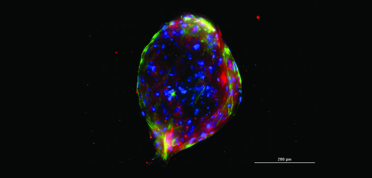
“Identification of functionally distinct sub-populations of human chondrocytes and defining cellular and molecular identity of cells within the so called ‘superficial zone’ of articular cartilage, the most vulnerable layer of cartilage degradation of which is implicated in the early stages of osteoarthritis.”
“We were also able to show that cells forming superficial zone can be generated from human pluripotent stem cells. Finally, we showed that transplantation of engineered cells into animal joints resulted in the generation of human cartilage and that this human tissue was fully integrated into recipient’s tissue fully restoring the superficial zone of damaged articular cartilage.”
Dr. Evseenko commented to OTW, “The current study defines a novel approach for generating functional articular chondrocytes from pluripotent stem cells with robust ability to restore the superficial zone. Systematic analysis of human skeletal development was critical for our understanding of how human joint forming cartilage is naturally formed. This knowledge was instrumental for making similar cells in vitro and using them therapeutically for articular cartilage tissue engineering.”
“Hopefully one day not too far in the future, they will be able to tell their patients with focal articular cartilage lesions, that their disease can be treated with novel cell-based therapies that will do more than just manage pain. This early stage intervention will be critical for prevention of articular cartilage degeneration and invasive procedures such as total joint replacement.”
React:
Discussion
This is a fascinating development. In my practice we've seen similar outcomes with the revised protocol. The key differentiator seems to be patient selection criteria. Has anyone else noticed the correlation with BMI thresholds?
Great point. I'd push back slightly on the conclusion, the sample size in the cited study is too small to draw population-level inferences. That said, the directional signal is compelling and worth a larger RCT.
We implemented a similar approach last year. Early results are promising but we're still gathering 12-month follow-up data. Happy to share our protocol if anyone is interested.
Join the conversation
Orthopedic professionals are discussing this. Sign in and upgrade to read every comment and add your voice.