Patients with missing bone due to injury or disease have a plethora of options for bone grafts to replace it. There’s autograft, or allograft, or any number of calcium-based products, recombinant bone morphogenic proteins (BMP) and even glass (BioGlass).
New Bone Grafting Option
2 min read Premium comments
Secondary#allograft#autograft#bonegrafts
Now researchers at New York University are developing another option: 3D printed ceramic implants that dissolve slowly within the body, stimulating bone to grow in their place. 3D printing allows scientists to create implants that precisely fit the defects. They can also be coated with chemicals that promote bone growth.
According to the researchers, the animals they tested were able to absorb the implants into their natural bone, healing defects.
“Our three-dimensional scaffold represents the best implant in development because of its ability to regenerate real bone,” said study senior investigator and biomedical engineer Paulo G. Coelho, D.D.S., Ph.D., the Dr. Leonard I. Linkow Professor at NYU Dentistry and a professor of plastic surgery at NYU School of Medicine.
“Our latest study results move us closer to clinical trials and potential bone implants for children living with skull deformations since birth, as well as for veterans seeking to repair damaged limbs.”
According to the researchers, their ceramic 3D printed implants closely resembles the shape and composition of real bone. They contain beta tricalcium phosphate, which is similar to components in natural bone, making the implants resorbable over time. They are coated with dipyridamole, a blood thinning agent that stimulates bone growth and attracts bone cells to the implant.
“Dipyridamole has proven to be key to the implant’s success,” said study co-investigator Bruce N. Cronstein, M.D., the Dr. Paul R. Esserman Professor of Medicine at NYU School of Medicine, who also serves as the director of the Clinical and Translational Science Institute, and chief of the Division of Translational Medicine at NYU Langone Health.
Cronstein perfected the drug’s use during device testing. “And because the implant is gradually resorbed, the drug is released a little at a time and locally into the bone, not into the whole body, thereby minimizing risks of abnormal bone growth, bleeding, or other side effects.”
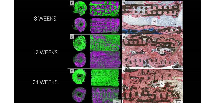
The researchers used the test scaffolds to repair small holes surgically made in the jaws of mice, as well as missing bone pieces as much as 1.2 centimeters long in rabbit limbs and jaws. On average, 77% of each scaffold was reabsorbed by the animals’ bodies six months after implantation.
CT scans of the implant sites showed almost no trace of beta tricalcium phosphate. Weight-bearing tests showed that the new bone was the same strength as the original undamaged bone.
Clinical trials are still several years away, according to the researchers, and the next step is to test the scaffolds in larger animals.
React:
Discussion
This is a fascinating development. In my practice we've seen similar outcomes with the revised protocol. The key differentiator seems to be patient selection criteria. Has anyone else noticed the correlation with BMI thresholds?
Great point. I'd push back slightly on the conclusion, the sample size in the cited study is too small to draw population-level inferences. That said, the directional signal is compelling and worth a larger RCT.
We implemented a similar approach last year. Early results are promising but we're still gathering 12-month follow-up data. Happy to share our protocol if anyone is interested.
Join the conversation
Orthopedic professionals are discussing this. Sign in and upgrade to read every comment and add your voice.