In a highly significant new study of 2,007 knees, UK researchers used compositional magnetic resonance imaging (MRI) to successfully detect early cartilage degeneration.
2,007 Knees Reveal Cartilage Degeneration Clues
2 min read Premium comments
#osteoarthritisSecondary#knee#articularcartilage
How did they do it and what did they learn?
The detailed answers are in their published work, “Systematic review and meta-analysis of the reliability and discriminative validity of cartilage compositional MRI in knee osteoarthritis,” which appears in the September 2018 edition of Osteoarthritis and Cartilage.
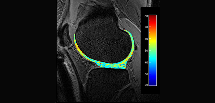
Co-author James MacKay, M.A., M.B., B.Chir. with the Department of Radiology at the University of Cambridge School of Clinical Medicine, explained, “’Compositional’ MRI techniques can detect changes in the composition of articular cartilage (e.g., loss of proteoglycans and/or collagen) before gross structural damage has occurred.”
“Lots of clinicians and researchers are interested in using them because they offer a non-invasive way to both identify early cartilage degeneration and monitor response to treatments aiming to improve cartilage health.”
“At the moment these techniques are mostly used in the research setting. We wanted to enable their more widespread use by establishing that they can provide reliable/reproducible results and are able to distinguish healthy from diseased cartilage. This was the impetus for us to perform our systematic review and meta-analysis of the literature on this topic.”
“I think there were two key results.
First, the majority of the compositional MRI techniques we evaluated demonstrated excellent reliability. This is very important if we want to use these methods as quantitative imaging biomarkers (i.e., objective measures of cartilage health).
Second, we showed that the two most commonly used compositional MRI techniques, T2 mapping and T1rho mapping, were able to discriminate between healthy cartilage and cartilage in people with osteoarthritis (OA)—this was true for both general OA and early OA populations.”
“At present I would caution against making routine clinical use of commercially available compositional MRI methods (e.g., T2 mapping) as there are still several outstanding issues to be addressed such as standardization of MRI protocols and analysis methodologies.”
“However, we are moving towards a time when I believe that these techniques could have real clinical impact.”
“Our paper will form part of an international collaborative effort to establish T1rho and T2 mapping as accepted quantitative imaging biomarkers via the Radiological Society of North America’s (RSNA) Quantitative Imaging Biomarkers Alliance (QIBA), which will include addressing many of these outstanding issues.”
“Cartilage compositional MRI techniques can detect changes in cartilage composition before gross structural changes occur. Such techniques are of potential use in detecting early degeneration and in monitoring changes in cartilage health over time. Our paper is part of a wider effort to enable the more widespread clinical use of cartilage compositional MRI.”
React:
Discussion
This is a fascinating development. In my practice we've seen similar outcomes with the revised protocol. The key differentiator seems to be patient selection criteria. Has anyone else noticed the correlation with BMI thresholds?
Great point. I'd push back slightly on the conclusion, the sample size in the cited study is too small to draw population-level inferences. That said, the directional signal is compelling and worth a larger RCT.
We implemented a similar approach last year. Early results are promising but we're still gathering 12-month follow-up data. Happy to share our protocol if anyone is interested.
Join the conversation
Orthopedic professionals are discussing this. Sign in and upgrade to read every comment and add your voice.