“Incorporation and Remodeling of Structural Allografts in Acetabular Reconstruction: Multiscale, Micro-Morphological Analysis of 13 Pelvic Explants,” appears in the August 15, 2018 edition of The Journal of Bone and Joint Surgery.
Acetabular Reconstruction: What Happens to Graft Bone Over Time?
2 min read Premium comments
Secondary#allograft#hip#acetabularreconstruction
Senior author Tim Rolvien, M.D., with the Department of Osteology and Biomechanics, University Medical Center Hamburg‐Eppendorf in Germany, told OTW, “While structural allografts are successfully used in revision total hip arthroplasty, the extent to which the graft bone is incorporated over time had not been analyzed.”
“In fact, previous studies were limited to only clinical data or very few cases. Due to our expertise in histological analysis of bones and our orthopedic background, we were excited to perform this multiscale analysis of structural allograft used in acetabular reconstruction.”
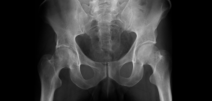
The authors wrote, “Thirteen acetabula were retrieved post mortem, and the incorporation properties of the bone allografts were analyzed using a hierarchical approach of imaging techniques including contact radiography, high-resolution peripheral quantitative computed tomography (HR-pQCT), histological analysis of undecalcified specimens, and quantitative backscattered electron imaging (qBEI).”
“The distance between the current allograft bone and host bone borders (i.e., current overlap) as well as the distance between the original allograft bone and host bone borders (i.e., total ingrowth) were assessed.”
According to Dr. Rolvien, they study showed that:
- A direct contact between the allograft and host bone was achieved in all cases along 25-100% of the interface;
- Allografts were successfully incorporated within the host bone, which could be measured by an overlap of the allograft and host bone border;
- The center of the allograft bone remained un-remodeled even in the case in which the allograft had been in situ for the most time;
- The allograft bone showed an intact trabecular structure and significantly higher mineralization compared with the host bone;
- Neither the time that the allograft was in situ nor the degree of contact between the host and allograft bone correlated with the overlap of the host and allograft bone.”
“From a histological point of view, structural allografts represent a very good option for the treatment of large acetabular bone defects. A tight junction between the host bone and the allograft bone is most likely a prerequisite for a successful remodeling process and consecutive incorporation of the allograft bone.”
“Our study on the incorporation of structural allografts not only included a large number of cases with long-term follow-up but also was based on the analysis of complete pelvic explants, in contrast to studies of biopsy specimens only, and therefore conclusively clarifies the extent of incorporation of structural allografts in acetabular reconstruction.”
“In other words, our study provides the first systematic multiscale evaluation of successfully implanted structural allografts and forms the scientific basis for their clinical use in revision THA.”
React:
Discussion
This is a fascinating development. In my practice we've seen similar outcomes with the revised protocol. The key differentiator seems to be patient selection criteria. Has anyone else noticed the correlation with BMI thresholds?
Great point. I'd push back slightly on the conclusion, the sample size in the cited study is too small to draw population-level inferences. That said, the directional signal is compelling and worth a larger RCT.
We implemented a similar approach last year. Early results are promising but we're still gathering 12-month follow-up data. Happy to share our protocol if anyone is interested.
Join the conversation
Orthopedic professionals are discussing this. Sign in and upgrade to read every comment and add your voice.