New work tackles the daunting task of illustrating fracture lines in complex cases.
Fracture Patterns in Triplane Fractures
2 min read Premium comments
Secondary#axialfracture#triplanefracture
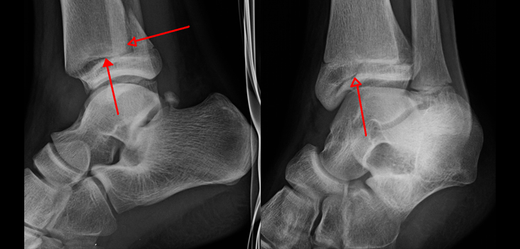
This important study: “Surgically Relevant Patterns in Triplane Fractures: A Mapping Study,” in the June 20, 2018 edition of The Journal of Bone and Joint Surgery, does the difficult work of illustrating fracture lines in the axial plane and bringing order to the multiple patterns of fractures.
According to the authors, “Triplane ankle fractures are complex transitional fractures that often necessitate surgical treatment. Axial fracture lines determine optimal screw trajectories for fixation. The purpose of our study was to identify fracture patterns in triplane fractures by illustrating fracture lines in the axial plane of the distal tibial metaphysis and epiphysis.”
“We retrospectively reviewed records of children presenting with ankle fractures at one center from January 2007 through June 2017.”
“Thirty-three cases of triplane fractures with available computed tomographic (CT) scans were identified. Fractures in the axial plane of the metaphysis were identified 10 mm proximal to the physis, and fractures in the epiphysis were identified midway between the physis and distal tibial articular surface.”
“Fracture lines were drawn and were superimposed on unfractured bone templates to generate fracture maps, and heat maps were then created to show areas of high and low fracture densities.”
“In the metaphysis, the most common fracture pattern was medial-lateral lines in the posterior metaphysis. This metaphyseal pattern was consistent across 2, 3, 4, and 5-part fractures. There were clear zones of rare fracture involvement in the anterior and anterolateral metaphysis.”
“In the epiphysis, anterior-to-posterior fracture lines were common in the anterior epiphysis. All cases had an epiphyseal fracture exit through the anterior epiphysis. Fracture extension into the posteromedial epiphysis was a feature of all fracture classes.”
“Fracture mapping of triplane fractures suggests consistent axial fracture patterns in the metaphysis and epiphysis with additional class-dependent fractures in the epiphysis. This study provides visual guidelines to assist surgeons in understanding the axial fracture patterns of individual triplane fractures for surgical planning.”
Co-author Paul Sponseller, M.D., M.B.A. told OTW, “This topic was of interest because we wanted to bring some order to the multiple patterns of triplane fractures which have been described. We found that, in fact there were very common/consistent themes for fracture lines at each axial level of the ankle.”
“This is important because it allows more minimally invasive options for fixation. It also allows more informed placement of any surgical incision. The best placement options for percutaneous fixation were described in the illustrations.”
React:
Discussion
This is a fascinating development. In my practice we've seen similar outcomes with the revised protocol. The key differentiator seems to be patient selection criteria. Has anyone else noticed the correlation with BMI thresholds?
Great point. I'd push back slightly on the conclusion, the sample size in the cited study is too small to draw population-level inferences. That said, the directional signal is compelling and worth a larger RCT.
We implemented a similar approach last year. Early results are promising but we're still gathering 12-month follow-up data. Happy to share our protocol if anyone is interested.
Join the conversation
Orthopedic professionals are discussing this. Sign in and upgrade to read every comment and add your voice.