A new technique from the UK could help physicians assess osteoarthritis (OA) on a whole new, detailed level. The researchers undertook a study titled, “A new quantitative 3D approach to imaging of structural joint disease.” The study appears in the June 18, 2018 edition of Scientific Reports.
New Algorithm Creates 3D Map From CT Data
2 min read Premium comments
Secondary#3dimaging#jointdisease#tomturmezei
Co-author Tom Turmezei, Ph.D. of the Cambridge University Engineering Department, Cambridge, UK, Department of Radiology, Norfolk and Norwich University Hospital, Norwich, UK, told OTW, “We are a collaborative group of engineers, radiologists and physicians based in Norwich and Cambridge, UK, that looks to solve clinical imaging problems with technical solutions.”
“At the outset, we knew that the ability to assess OA with 2D radiographs was not good enough to detect subtle changes that could have important implications for patients, doctors, and researchers, so we decided to develop a new algorithm for the structural assessment of joints in 3D using computed tomography (CT) imaging data.”
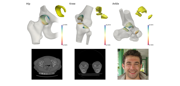
“Since this was a technical validation study, we showed that the technique worked, and was able to map joint space width (the distance between bones at a joint that is a surrogate marker for osteoarthritis) in 3D accurately to the nearest tenth of a mm.”
“The technique also has potential to be twice as sensitive as radiographs in detecting subtle changes in joint space width of 0.2 mm compared to 0.45 mm as the reported best from radiographs. Finally, we also showed that the technique was very robust, working at a number of different joints, including the hip, knee, and ankle.”
“We now know that we can use clinical CT scans to map an important marker of osteoarthritis in 3D across a joint, which could identify patients with disease earlier than the current gold standard radiographic method. As a result, this could allow important interventions to be started before the joint fails, such as lifestyle change and physiotherapy.”
“This new method should also allow researchers to spot whether new therapies in development are effective in a realistic timescale for clinical trials, something that has not been possible using radiographs.”
“The ability to map joint space width in 3D from clinical CT data is a sensitive, reliable and accurate technique that has the potential to change our understanding of osteoarthritis and how patients progress to joint failure.”
“We are now performing further research to show how the joint space is different in individuals without radiographic signs of disease that progress to total hip replacement, therefore revealing the potential to identify at risk patients even earlier. 3D joint space maps are also easier to interpret and show how a joint is failing and could even be used to explain to patients how and exactly where their osteoarthritis is progressing.”
React:
Discussion
This is a fascinating development. In my practice we've seen similar outcomes with the revised protocol. The key differentiator seems to be patient selection criteria. Has anyone else noticed the correlation with BMI thresholds?
Great point. I'd push back slightly on the conclusion, the sample size in the cited study is too small to draw population-level inferences. That said, the directional signal is compelling and worth a larger RCT.
We implemented a similar approach last year. Early results are promising but we're still gathering 12-month follow-up data. Happy to share our protocol if anyone is interested.
Join the conversation
Orthopedic professionals are discussing this. Sign in and upgrade to read every comment and add your voice.