Swedish researchers have developed a new biomedical adhesive that can be used for fracture fixation. Their work, “High‐Performance Thiol–Ene Composites Unveil a New Era of Adhesives Suited for Bone Repair,” was published in the April 2018 edition of Advanced Functional Materials.
What! No Elmer’s? Novel Fracture Adhesive in Lab
2 min read Premium comments
Secondary#fracture#knee#boneglue
Michael Malkoch, Ph.D., professor in Functional Organic Nanomaterials and a senior researcher in Biomedical Engineering at the KTH Royal Institute of Technology in Stockholm, Sweden, and co-author on the study, told OTW, “The journey was initiated by colleagues from Karolinska University Hospital (neurosurgeons) and then after at South Stockholm Hospital (hand surgeons).”
“However, for use of biomedical adhesive in clinical setting a great number of criteria need be fulfilled. For soft-tissue fixations, medically approved super glues (e.g., Histoacryl) has extensively been used. Unfortunately, superglues are extremely reactive and do not discriminate between tissues resulting in adhesion between soft-tissue, tendons, etc. Additionally, super glues have low viscosity and [are] quite fragile in nature. Low viscosity may lead to diffusion into the fracture and thereof make more harm than good. As they are fragile they can’t really appropriately fixate fracture for proper healing.”
“Dental adhesives on the other hand have great fixation power but are based on typically questionable acrylate systems with bisphenol A as an important component (as well as a large portion of glass filler).
For adhesives to realistically be used for fracture fixations it was necessary to design novel glue components that can via robust and selective chemistry (click chemistry) transform to hard adhesives with unprecedented adhesion to wet bone tissue.”
“In this study we showcased a feasible approach to fixate bone fracture using the fiber reinforced adhesive patch (FRAP) method.”
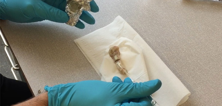
“The setup is simple and has great similarity to the one used in dentistry, i.e., a bottom-up build-up of a customized FRAP for a given fracture. We used porcine metacarpals as they were most resembling human fingers and we utilized both transversal and oblique fractures. The FRAP is built by first cleansing the bone surface from soft tissue followed by careful molt of the bone surface. A primer was then applied for efficient bonding to the bone. A two-component adhesive with hydroxyapatite fillers was then added and that efficiently binds to the primer, chemically.”
“Biomedical approved fiber mesh was imbedded and the procedure can be repeated all over again depending on the load that the FRAP need to withstand. A top coating of the two-component adhesive completes the FRAP build-up. The FRAP is cured using a high-energy visible light source typically used by dentists.”
“While we have focused on finger fractures, it is inevitable that these adhesives are suited for the larger community of orthopedics (cranial, ankle, wrist etc.). The strategy can easily be transferred to minimally invasive surgery (local anesthesia) and towards more sensitive tissues, i.e., cervical fractures. Importantly, the FRAPs can coexist with current implants, especially when shattered fractures need proper fixation. Not to forget, the topological fixation strategy makes drilling, for instance in elderly patients, obsolete.”
React:
Discussion
This is a fascinating development. In my practice we've seen similar outcomes with the revised protocol. The key differentiator seems to be patient selection criteria. Has anyone else noticed the correlation with BMI thresholds?
Great point. I'd push back slightly on the conclusion, the sample size in the cited study is too small to draw population-level inferences. That said, the directional signal is compelling and worth a larger RCT.
We implemented a similar approach last year. Early results are promising but we're still gathering 12-month follow-up data. Happy to share our protocol if anyone is interested.
Join the conversation
Orthopedic professionals are discussing this. Sign in and upgrade to read every comment and add your voice.