Organ transplants and other advanced tissue treatments face a seemingly impassable bottleneck. There are only a finite number of organ donors or other sources of biomaterials. Even in the best of cases the organs and tissues which have been donated are rarely perfectly compatible with the recipient and thus may not work well.
Bioprinter Designed to Print Body Parts
2 min read Premium comments
Secondary#bioprinter#bodyparts
“Bioengineers would like to be able to bypass conventional sources altogether and grow organs and tissues in the lab. This would not only provide the medical community with an unlimited supply of healthy, sterile materials, but would also allow doctors and surgeons to have biomaterials made to their specifications.”
“The trouble is, living tissue is incredibly complex and contains many different kinds of cells, blood vessels, nerves, and mechanical structures.”
One way to proceed is to “build a scaffold out of biocompatible material like the hydrogels poly (ethylene glycol) diacrylate (PEGDA) and gelatin methacryloyl (GelMA). A scaffold made of these materials which mimic the structure of living tissue, acts like the cartilage in an infant’s body. When first born, much of a human baby’s skeleton is cartilage, but as it grows and matures, this is replaced by bone tissue. In the artificial tissue, stem cells are introduced that grow into the scaffolding and replace it.”
“One technique for creating these scaffolds is called stereolithography. This is a light-based process where hydrogel mixed with stem cells is laid down by a 3D printer while at the same time a beam of light causes molecular bonds to form, hardening the gel.”
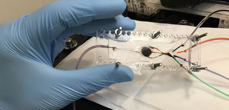
The UCLA bioprinter designed by a team lead by bioengineer Ali Khademhosseini, Ph.D. is based on this technique. It also incorporates a microfluidic chip about the size and shape of a microchip. This can print with more than one cell-infused material at a time and is joined by a digital micromirror made of a million mirrors that move independently of one another.
According to UCLA, the automated mirrors create a pattern for each layer of the object being printed while the light cures the gel.
“So far, the printer has been used to create simple shapes, 3D simulations of muscle tissue and muscle-skeleton connective tissues, as well as fake tumors complete with blood vessels. …the structures have been implanted in rats without being rejected.”
“Tissues are wonderfully complex structures, so to engineer artificial versions of them that function properly, we have to recreate their complexity,” says Khademhosseini. “Our new approach offers a way to build complex biocompatible structures made from different materials.”
The research, titled “Microfluidics‐Enabled Multimaterial Maskless Stereolithographic Bioprinting,” is published in Advanced Materials.
React:
Discussion
This is a fascinating development. In my practice we've seen similar outcomes with the revised protocol. The key differentiator seems to be patient selection criteria. Has anyone else noticed the correlation with BMI thresholds?
Great point. I'd push back slightly on the conclusion, the sample size in the cited study is too small to draw population-level inferences. That said, the directional signal is compelling and worth a larger RCT.
We implemented a similar approach last year. Early results are promising but we're still gathering 12-month follow-up data. Happy to share our protocol if anyone is interested.
Join the conversation
Orthopedic professionals are discussing this. Sign in and upgrade to read every comment and add your voice.