The FDA 3D-printed interbody fusion cage clearances keep coming.
Stryker 3D-Printed Lumbar Cage Cleared
2 min read Premium comments
#stryker#backpainSecondary#degenerativediscdisease#fusion
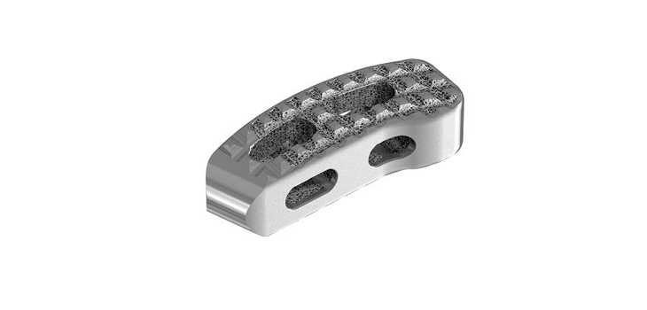
On March 7, 2018, Stryker Spine announced its Tritanium TL Curved Posterior Lumbar Cage received 510(k) clearance from the FDA. The cage is intended for use as an aid in lumbar fixation.
The company says the cage is a hollow implant consisting of a “unique configuration of both solid and porous structures, which are simultaneously built using” the company’s proprietary 3D printing process called, AMagine. The technology, according to the company, is “deliberately designed for fusion and inspired by the microstructure of cancellous bone.”
Tritanium Technology
The cage features Stryker’s Tritanium Technology, a “novel, highly porous titanium material designed for bone ingrowth and biological fixation. This In-Growth technology has demonstrated that osteoblasts (bone cells) infiltrated, attached to, and proliferated on the porosity of the Tritanium technology. The unique porous structure is designed to create a favorable environment for cell attachment.”
In addition, the company says the tritanium material “may be able to wick or retain fluid in comparison to traditional titanium material. The TL cage complements the Tritanium PL cage, and together they offer alternative posterior lumbar solutions for spinal surgeons.”
Stryker’s Spine President Brad Paddock said “…the cage is accompanied by a new Anterior Placement System that is designed for versatility and procedural flexibility. From instrumentation ergonomics and visualization, to a simplified technique with tactile feedback, Tritanium TL’s Anterior Placement System and cage design redefine implant steerability for surgeons.”
To be made available to surgeons in the second quarter of 2018, “…the cage features open central graft windows and lateral windows to help reduce stiffness of the cage, aid in visualization of fusion, and allow for bone graft containment.” The company said both cages are “shaped for steerability, its multidirectional teeth are designed for multidirectional fixation as the cages can be steered and rotated to the surgeon’s desired placement.”
They are also both designed to “maximize surface area” for endplate contact with the implant. The TL cage, says the company, “has a smooth, tapered leading edge to facilitate insertion into the intervertebral space and a central column spanning endplate to endplate for structural integrity.”
The TL Cage will be available in a broad range of footprints, heights, and lordotic angles.
Indications
The company states the device is “indicated for use with autograft and/or allogenic bone graft comprised of cancellous and/or corticocancellous bone graft when used as an adjunct to fusion in patients with degenerative disc disease (DDD) at one level or two contiguous levels from L2 to S1…and is to be implanted via a posterior approach.”
React:
Discussion
This is a fascinating development. In my practice we've seen similar outcomes with the revised protocol. The key differentiator seems to be patient selection criteria. Has anyone else noticed the correlation with BMI thresholds?
Great point. I'd push back slightly on the conclusion, the sample size in the cited study is too small to draw population-level inferences. That said, the directional signal is compelling and worth a larger RCT.
We implemented a similar approach last year. Early results are promising but we're still gathering 12-month follow-up data. Happy to share our protocol if anyone is interested.
Join the conversation
Orthopedic professionals are discussing this. Sign in and upgrade to read every comment and add your voice.