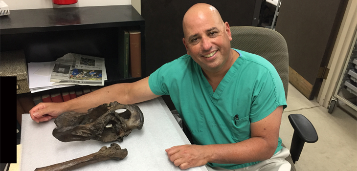
Computed Tomography (CT) technology is used every day by orthopedic surgeons like Robert Klapper, M.D., co-director of the Joint Replacement Program at Cedars-Sinai Medical Center in Los Angeles, California, to treat patients with bone fractures and worn out joints, but recently Klapper discovered that this technology can also be used to understand the prehistoric world of saber-toothed cats.
Cedars-Sinai Surgeon Uses CT Scan to Get Old Bones to Talk
2 min read Premium comments
Secondary#hipdysplasia#computedtomographyscans#pelvic
Saber-toothed cats have been extinct for more than 12,000 years and little is known about them. One of the biggest debates among paleontologists has been whether they were solitary hunters or hunted in packs.
According to a press release, Klapper is working with the paleontologists at the La Brea Tar Pits and Museum also in Los Angeles to use Cedars-Sinai’s most advanced CT scan machines to study the pelvic and femur bones of the extinct cat in the hopes it shed some light on how saber-toothed cats lived and roamed.
“The most modern technology allowed these bones to speak to us, and they had a lot to say,” Klapper said.
He told OTW that he first became interested in prehistoric fossils about 20 years ago when while visiting the museum he noticed that all the dire wolf skulls on a wall looked alike.
“It was overwhelming how similar they looked and I thought where are the abnormalities in the bones? There were no signs of fractures or cancer or arthritis so I bugged the security officer until he let me talk to the collections manager at the time, Chris Shaw, who took me into the bowels of the museum to get a closer look at some of the bones.”
Shaw pulled out the bones from one particular saber-toothed cat that the paleontologists had a lot of questions about. They had hypothesized that the cat died from an infection, but Klapper noticed immediately signs of arthritis, one side of the pelvis, the acetabulum, was a smooth surface but the other side looked like a hand grenade had gone off.
Klapper got permission to take the bones of a 12,000 year old saber-toothed cat to Cedar Sinai for scanning and while he completed the scanning at that time, he didn’t really study the images closely until more recently. He said that life just got real busy with his practice and his health and sports radio show Weekend Warrior on ESPN.
The scans it turns out could change the way we view how the saber-toothed cat lived and hunted because they showed that the cat in question had hip dysplasia, an abnormal development of the hip joint. The fact that this cat was able to grow into adulthood despite having a constant limp slowing it down indicates that it lived in a pack because hunting on its own it most likely wouldn’t have survived as long as it did.
Klapper believes that the CT scans of the cat’s bones will also help us better understand how the ball and socket deform each other and can be used to create a prosthetic hip joint for the tallest human patients with dysplasia. Currently there are no prosthesis available for patients who are 7 feet tall.
Emily Lindsey, Ph.D., a paleoecologist and assistant curator and excavation site director at La Brea Tar Pits, told OTW, “We are really excited about the collaboration with Dr. Klapper and we hope to continue the partnership to look at other specimens.”
Klapper is currently having 3D models of the CT scans made, and the museum is exploring putting together an exhibit of the findings.
React:
Discussion
This is a fascinating development. In my practice we've seen similar outcomes with the revised protocol. The key differentiator seems to be patient selection criteria. Has anyone else noticed the correlation with BMI thresholds?
Great point. I'd push back slightly on the conclusion, the sample size in the cited study is too small to draw population-level inferences. That said, the directional signal is compelling and worth a larger RCT.
We implemented a similar approach last year. Early results are promising but we're still gathering 12-month follow-up data. Happy to share our protocol if anyone is interested.
Join the conversation
Orthopedic professionals are discussing this. Sign in and upgrade to read every comment and add your voice.