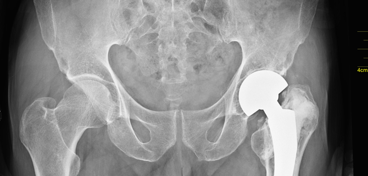
According to a new study, digital imaging can be effectively applied during total hip arthroplasty (THA). The study, “Digital Radiography in Total Hip Arthroplasty: Technique and Radiographic Results,” appears in the February 7, 2018 edition of The Journal of Bone and Joint Surgery.
Hip Surgery? Try Digital Imaging Intraoperatively
2 min read Premium comments
Secondary#hipsurgery#hip#digitalimaging
Co-author on the study, Brad L. Penenberg, M.D. with the Department of Orthopaedic Surgery at Cedars-Sinai Medical Center in Los Angeles, told OTW, “The availability of digital radiography has created a paradigm shift in image acquisition, image quality, and image analysis. I believe that this paper confirms for the first time, that by applying digital imaging technology it is possible to acquire a high-quality AP pelvis X-ray with a patient in the lateral position during THA.”
“It also confirms that this X-ray can be used to acquire the same information during the operation that we used to acquire only after surgery. In the intra-operative setting, however, we have an opportunity to make adjustments that have the potential to lead to a new level of accuracy in any THA.”
“In the past, using traditional instruments, bony landmarks, and an assumed pelvic position, it was not unusual to see cup position out of the target range. We saw femoral components on the post op X-ray that were undersized and in varus when we thought they were as tight a fit as possible based on ‘feel.’”
“Limb length and offset assessment was imprecise. Dislocation and limb length inequality are among the most common subjects of malpractice cases following THA.”
“They are often related since a common solution for impingement during THA is to change to a longer head. The intra-op radiograph can identify that perhaps the cup position should be changed rather than unnecessarily adding to limb length. We also saw the occasional unseated cup or an unintended screw trajectory.”
“A high quality, well-oriented intra-op X-ray has the potential to minimize, and hopefully eliminate, all of those outliers.”
“It is possible to obtain a reliable AP pelvis X-ray during THA with the patient in the lateral position. Relevant bony landmarks can be identified and utilized for the necessary measurements.”
“Once we established image accessibility and quality, we were able to improve efficiency of image analysis by applying automated landmark annotation software (Radlink).”
“The surgeon, while scrubbed, can do his own measurements by simply touching points and making minor line adjustments. The specially designed software walks the surgeon through the necessary steps so that, unlike the use of traditional PACS tools, the process of measurement can take only a few seconds.”
The image analysis software (Radlink) can be used with any portable X-ray unit or C-arm.”
React:
Discussion
This is a fascinating development. In my practice we've seen similar outcomes with the revised protocol. The key differentiator seems to be patient selection criteria. Has anyone else noticed the correlation with BMI thresholds?
Great point. I'd push back slightly on the conclusion, the sample size in the cited study is too small to draw population-level inferences. That said, the directional signal is compelling and worth a larger RCT.
We implemented a similar approach last year. Early results are promising but we're still gathering 12-month follow-up data. Happy to share our protocol if anyone is interested.
Join the conversation
Orthopedic professionals are discussing this. Sign in and upgrade to read every comment and add your voice.