Study: When Spine Images Don’t Match Symptoms
New Spine Study: When Images Don’t Match Symptoms; New Tobramycin Study; Machine Learning Transforms Meniscal Imaging
7 min read Premium comments
#lumbarspine#orthopedics#chronicbackpain#facetjointinfiltration#meniscaldamage#surgicalwoundcontamination
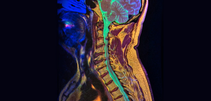
Researchers from the UK are stressing that there is no gold standard for diagnosis and the related indication for surgery in chronic lumbar back pain patients. Their study, “Predictability of the effects of facet joint infiltration in the degenerate lumbar spine when assessing MRI scans,” appears in the November 21, 2017 edition of the Journal of Orthopaedic Surgery and Research.
Ulf Krister Hofmann, M.D., orthopedic surgeon with University Hospital of Tübingen in Germany and co-author on the study, told OTW, “Due to advances in surgical techniques and perioperative management, spine surgery has seen great improvements over the past three decades. This is a blessing for many patients suffering from chronic lumbar back pain who in many cases today can be treated successfully by surgery.”
“The danger of being so successful with surgery is, that when your only tool available is a hammer, every problem tends to look a bit like a nail.”
“To identify patients who will actually benefit from surgery remains challenging. There are also many conditions that can manifest themselves as chronic lumbar back pain, and that can not be addressed by surgery, such as psychiatric disorders like depression, a nonspecific functional instead of a physical etiology, or referred pain from extraspinal causes (e.g., ovarian cyst, pancreatitis, ulcer).”
“In many patients, clinical history and symptoms and radiologic findings are conclusive, which makes the decision-making process easier. There are, however, also these patients where quite a discrepancy can be observed between their clinical presentation and the morphologic changes present in MRI or CT.”
“Like laboratory tests or histopathological findings, imaging results are often considered to be solid evidence by physicians and patients. The confidence in these imaging findings with respect to their ability to predict a painful condition appears however, somewhat anticipated given the available data in the literature.”
“In our centre of orthopaedic surgery we see many patients with a discrepancy between clinical and radiological findings.”
“It is in these patients that we additionally perform image-guided local analgesic or anti-inflammatory infiltrations at possible sites of pain generation to temporarily simulate the effect of surgery.”
“Such possible sites are, for example, the facet or sacroiliac joints, the epidural space, the deep back muscles, or the spinal nerves at their exit through the intervertebral foramen, as well as the hip as a differential diagnosis for chronic lumbar back pain.”
“From the achieved improvement reported by the patient, the specialist can draw further conclusions as to the cause of the patient’s symptoms. This allows to better identify patients who might and who might not benefit from surgery, and to define the actual scope of the planned surgery.”
“Reimbursement of these infiltrations can, however, be tedious since many insurance companies do not appreciate the diagnostic value of these procedures before surgery.”
“We, therefore, wanted to evaluate the ability of modern 3 tesla MRI to predict reported pain relief after facet joint infiltration in patients with chronic lumbar back pain. We hypothesized that, as pathological grading increased in MRI scans, pain alleviation would also increase after bilateral facet joint infiltration.”
“In our study we graded 50 MRI scans of patients with chronic lumbar back pain using a wide range of classification and measurement systems. The reported effect of facet joint injections at the site was recorded, and a comparative analysis performed.”
“When we allocated patients according to their reported pain relief, 27 showed no improvement (0–30% improvement on the NRS), 16 reported good improvement (31–75%) and 7 reported excellent improvement (> 75%).
“MRI features assessed in this study did, however, not show any relevant correlation with reported pain relief after facet joint infiltration.”
“Although we did not expect perfect agreement between reported pain relief and imaging findings, we were surprised to see this total lack of correlation between these two modalities!”
“If you do not assume that one of these two modalities is completely meaningless, our results can only mean, that the information provided by infiltrations and imaging are complementary.”
“We do need to point out that the patients analysed in this study were all part of the cohort of patients, where clinical presentation and imaging findings did not match. It is likely, that in a group of patients with matching symptoms and MRI or CT findings the results would have been different.”
“Our investigated collective is, however, a relevant subset of those patients presenting with chronic lumbar back pain and it is usually these patients where it is such a challenge to identify the best treatment strategy.”
“Specialists are accustomed to having some ‘gold standard’ they can refer to in their decision-making process. It is important to understand that there is no such gold standard for formulating a diagnosis and a resulting indication for surgery in chronic lumbar back pain patients.”
“Each modality—such as thorough clinical history, clinical examination, imaging techniques and local targeted infiltrations—has its strengths but also its flaws. It is the critical synopsis of all results obtained that allows one to identify the best treatment strategy for each patient.”
“To refer to the analogy mentioned above: only if you have all the tools in your toolbox available can you clearly see which problem is a nail, and which one isn’t.” — EH
Study: Intra-Wound Tobramycin Shows Promise
Researchers from Columbia University Medical Center, The Spine Hospital at New York Presbyterian have completed research in a rabbit model indicating that tobramycin does a good job of eradicating Escherichia coli (E. Coli). Their work, “Intrawound Tobramycin Powder Eradicates Surgical Wound Contamination: An In Vivo Rabbit Study,” appears in the December 15, 2017 edition of Spine.
Co-author Daniel Riew, M.D., co-chief of the spine division and director of cervical spine surgery at The Spine Hospital, commented to OTW, “We had previously published a study using a similar model and using vancomycin powder. While vancomycin is great for gram-positive organisms, it doesn’t work for gram-negatives such as E. Coli. So we thought we could do this study to determine if local tobramycin could eradicate E. Coli in a contaminated wound model.”
“We inoculated rabbits who had undergone a laminectomy and implantation of a titanium wire with E.Coli; 10 rabbits got intra-wound tobra and 10 did not. They were sacrificed on post-operative date #4. None of the rabbits who got tobra were infected whereas all of the ones without tobra got infected; 39 out of 40 culture specimens in the control and none out of 40 in the tobra group grew E. Coli.”
“This is a preliminary study and it only tells us that tobramycin is effective at eradicating E. Coli in rabbits. So, I cannot make recommendation for clinical usage yet. We are doing further studies regarding toxicity and dose. In the meantime, we do know that tobramycin impregnated cement does a nice job of treating wound infections and is not toxic. So, based on that information, I personally have used 70-140mg of intra-wound tobramycin when I was concerned about a gram-negative surgical contaminant.”
Dr. Riew concludes, “Stay tuned for more studies on this topic by our team.”
Machine Learning Transforms Meniscal Imaging
In the assessment of osteoarthritis (OA) progression, says new research from the UK, it would help to utilize 3D imaging to determine which meniscal pathologies undergo the most change. The study, “Where does meniscal damage progress most rapidly? An analysis using three-dimensional shape models on data from the Osteoarthritis Initiative,” is published in the January 2018 edition of Osteoarthritis and Cartilage.
Philip Conaghan M.B.B.S. Ph.D., professor of musculoskeletal medicine at the University of Leeds and deputy director of the National Institute for Health Research Leeds Biomedical Research Centre and co-author of the study, told OTW, “The meniscus is an integral part of the osteoarthritis process but much less studied than cartilage or even subchondral bone. In part this has been because it’s difficult to visualize even with MRI, which relies on a reader looking at a sequence of 2D images and which often fails to adequately show its 3-dimensional appearance.”
“This study included MRI images from people with definite OA and careful manual segmentation of the menisci in these images—but with the added value of supervised machine learning to enable correct placement of the menisci relevant to the tibia. And we were able to examine a number of different meniscal shapes as they degenerate over time.”
The authors wrote, “Knee images were selected from the progression cohort of the Osteoarthritis Initiative choosing participants with risk factors for medial OA progression. Medial and lateral menisci were manually segmented then analysed using a statistical shape model of the tibia as a reference surface.”
“Responsiveness was assessed at 1 year using standardized response means (SRMs) for four constructs: meniscal volume, extrusion volume, thickness and tibial coverage; anatomical sub-regions of these constructs were also explored. Paired images from 86 participants (median age 61.5, 49% female, 56% obese) were included.”
According to Dr. Conaghan, “Posterior medial meniscus was the location of most pathology. Despite relatively small numbers for a 12-month OA follow-up, there was responsiveness demonstrated for two of the meniscal measures.”
“There has been little investment in the OA field because demonstration of progression has been difficult in feasible time frames.”
“Modern image analysis has already provided the most responsive measures of OA progression in clinical trials, using either cartilage thickness or bone shape. This study provides a third responsive measure, meniscal shape. And it may add to the other measures.”
“This work underpins the benefits of modern 3D image analysis, which is not only being used for pre-joint replacement planning, but for understanding the importance of specific OA pathologies. In future we will be able to look at the range of meniscal pathologies and their relationship with symptoms.”
React:
Discussion
This is a fascinating development. In my practice we've seen similar outcomes with the revised protocol. The key differentiator seems to be patient selection criteria. Has anyone else noticed the correlation with BMI thresholds?
Great point. I'd push back slightly on the conclusion, the sample size in the cited study is too small to draw population-level inferences. That said, the directional signal is compelling and worth a larger RCT.
We implemented a similar approach last year. Early results are promising but we're still gathering 12-month follow-up data. Happy to share our protocol if anyone is interested.
Join the conversation
Orthopedic professionals are discussing this. Sign in and upgrade to read every comment and add your voice.