This week, Rochester, New York-based Carestream Health is demonstrating its new optional advanced metal artifact reduction software for its CARESTREAM OnSight 3D Extremity System at the Radiological Society of North America tradeshow.
Carestream Showcases Metal Artifact Reduction Software
1 min read Premium comments
Secondary
“Carestream’s second generation of software takes our state-of-the-art original metal reduction software to a new level. It provides enhanced flexibility depending on the metal content present and reduces the visual distortion caused by screws, implants, rods and other metal objects to create improved visibility and diagnostic confidence,” said Helen Titus, Carestream’s Worldwide Marketing Director for Ultrasound & CT.
“An intuitive touch screen interface allows technologists to adjust for either moderate or complex metal content,” says the company’s news release. “The metal artifact reduction software can be activated prior to the scan or it can be applied after the original reconstruction is complete. Both the original and corrected images are always available to view and compare.”
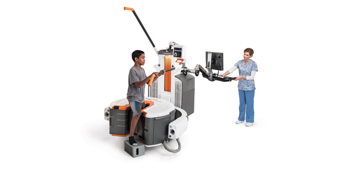
“The OnSight 3D Extremity System also assists surgeons in detecting occult and non-union bone fractures. Unlike traditional CT systems, this cone beam CT system has a large-area detector that captures a 3D image of the extremity in a single rotation, which takes only 25 seconds. A patient simply places the injured extremity into a donut-shaped opening in the system. Since the patient’s head and body are not confined, patients do not experience the claustrophobia that often occurs with traditional CT systems. Dose is significantly reduced because only the affected body part is imaged.”
Helen Titus told OTW, “The amount of metal content in an image can vary greatly. So we designed algorithms that accurately display a variety of metal devices as well as the surrounding tissue within an area of interest. We created two algorithms—one for anatomy with moderate metal content and the other for anatomy with complex metal content. This is designed to optimize visualization. And both the original image and corrected image are always available for viewing and comparison.”
React:
Discussion
This is a fascinating development. In my practice we've seen similar outcomes with the revised protocol. The key differentiator seems to be patient selection criteria. Has anyone else noticed the correlation with BMI thresholds?
Great point. I'd push back slightly on the conclusion, the sample size in the cited study is too small to draw population-level inferences. That said, the directional signal is compelling and worth a larger RCT.
We implemented a similar approach last year. Early results are promising but we're still gathering 12-month follow-up data. Happy to share our protocol if anyone is interested.
Join the conversation
Orthopedic professionals are discussing this. Sign in and upgrade to read every comment and add your voice.