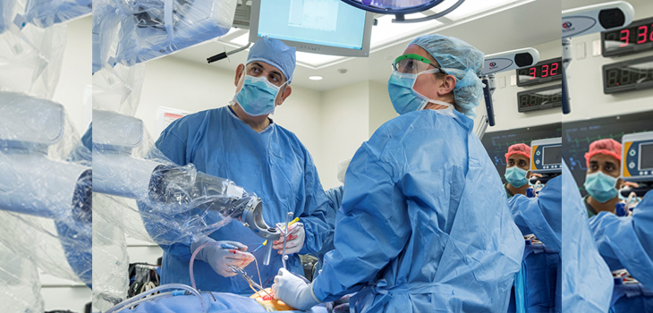
A system invented by a surgeon at The Johns Hopkins Hospital uses a real-time, image-guided robot to insert pedicle screws into a patient’s spine.
Johns Hopkins’ Robot to Insert Pedicle Screws
2 min read Premium comments
Secondary
“We are really excited to be able to offer this to our patients,” says Nicholas Theodore, M.D., professor of neurosurgery at the Johns Hopkins University School of Medicine and director of the Neurosurgical Spine Center of Johns Hopkins Medicine, in the October 10, 2017 news release.
Dr. Theodore invented the robot before joining the faculty at Johns Hopkins and maintains a financial interest in the technology. He added, “This will take what we neurosurgeons do on a daily basis, elevate the art, enable us to do things much more precisely and allow us to perform our best every day.”
As Johns Hopkins wrote in its news release, “When one drives a car and takes a quick glance to the side, often the steering wheel drifts in the same direction as the driver’s eyes. Theodore says current image-guided surgical procedures require the surgeon to look back and forth between the patient and an image, which causes imperfection of screw placement. While oftentimes these placements are ‘good enough,’ this wasn’t good enough for Theodore.”
“This new robot ‘marries’ a CT [computed tomography] scan of the patient with the actual patient, allowing the surgeon to point to a spot on the CT scan and tell the robot to aim for that same spot. Connected to a camera, which itself reads landmarks on the patient, the robot is able to process what the camera “sees” with the CT image in real time. Says Theodore, the biggest fear in this type of procedure is movement—what if the patient breathes or otherwise moves slightly—but this robot can sense changes in position and adjust accordingly.”
Dr. Theodore told OTW, “It was most interesting that after having built the first prototype that we were able to actually pick a point or trajectory on the spine and have the robot takes us precisely to that point in a quick, reproducible fashion.”
“Orthopedic surgeons need to know that this robotic platform can be accommodated to their workflow. That is to say that it can be used with a preoperative CT, an intraoperative CT or even fluoroscopy done during the case and use these medical images, once linked to the patient, to control the robots actions and provide real-time feedback as to the location of the end effector.”
React:
Discussion
This is a fascinating development. In my practice we've seen similar outcomes with the revised protocol. The key differentiator seems to be patient selection criteria. Has anyone else noticed the correlation with BMI thresholds?
Great point. I'd push back slightly on the conclusion, the sample size in the cited study is too small to draw population-level inferences. That said, the directional signal is compelling and worth a larger RCT.
We implemented a similar approach last year. Early results are promising but we're still gathering 12-month follow-up data. Happy to share our protocol if anyone is interested.
Join the conversation
Orthopedic professionals are discussing this. Sign in and upgrade to read every comment and add your voice.