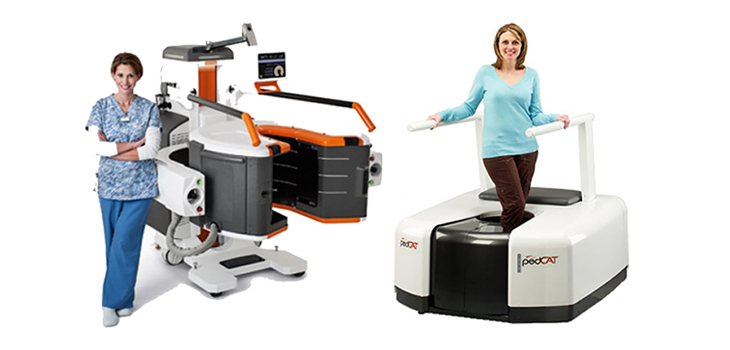
CurveBeam LLC and Carestream Health have announced that they will work together to support and facilitate education and research for weight-bearing computed tomography (CT) imaging. CurveBeam is located in Warrington, Pennsylvania, and Carestream Health is based in Rochester, New York.
CurveBeam, Carestream Collaborate for Weight-Bearing CT
2 min read Premium comments
Secondary
“The ability to capture weight-bearing CT images allows foot and ankle specialists to study foot biomechanics in three dimensions. Significant clinical research already has demonstrated weight-bearing CT has the potential to be the new standard of care for an initial diagnosis. However, much work remains to be done to increase awareness among orthopaedic specialists,” said Vinti Singh, CurveBeam marketing manager, in the June 6, 2017 news release. “Through collaboration, CurveBeam and Carestream can best support these efforts.”
“The image quality and detail captured with 3D weight-bearing CT exams allow the surgeon to view and measure anatomical abnormalities of lower extremities under natural load conditions,” said Helen Titus, Carestream’s worldwide marketing director for Ultrasound & CT. “The goal of this collaboration is to spread the word about this new imaging modality among orthopaedic specialists.”
The companies are supporting the Weight-Bearing CT International Study Group, a brain trust of orthopedic researchers that is striving to create standardized protocols for weight-bearing CT measurements and analysis. Their initial effort is the co-sponsoring of an evening scientific session during the American Orthopaedic Foot & Ankle Society (AOFAS) annual meeting in Seattle, Washington, July 12-15, 2017.
Helen Titus told OTW, “The image quality and detail captured with 3D weight-bearing CT exams allow surgeons to view and measure anatomical abnormalities of lower extremities under natural load conditions. A good example is patients with patellofemoral instability. Two of the challenges of this condition are identifying the cause of patella subluxation or dislocation, and then calculating the amount of correction that would be necessary when tibial tubercle transfer surgery is planned.”
“UBMD [The University at Buffalo, The State University of New York] Orthopaedics and Sports Medicine and Carestream Health are conducting an institutional IRB-approved clinical study to compare measures of tibial tubercle-trochlear groove offset (TT-TG) obtained on a conventional CT scanner to those obtained on the Carestream OnSight 3D Extremity System while the patient is standing, the quadriceps is active, and the knee is flexed to 30 degrees.”
Titus commented to OTW, “I anticipate that questions at the AOFAS evening scientific session will involve the clinical value of using weight-bearing CT exams and how images that show anatomy under the patient’s natural load aid the surgeon and radiologist in determining the correct treatment path or healing time. “I would also anticipate questions about how surgeons can use the 3D volume information for automatic measurements of joint spacing and other anatomical details. There may also be questions about patient workflow, reimbursement, and economic value of utilizing an in-office system.”
React:
Discussion
This is a fascinating development. In my practice we've seen similar outcomes with the revised protocol. The key differentiator seems to be patient selection criteria. Has anyone else noticed the correlation with BMI thresholds?
Great point. I'd push back slightly on the conclusion, the sample size in the cited study is too small to draw population-level inferences. That said, the directional signal is compelling and worth a larger RCT.
We implemented a similar approach last year. Early results are promising but we're still gathering 12-month follow-up data. Happy to share our protocol if anyone is interested.
Join the conversation
Orthopedic professionals are discussing this. Sign in and upgrade to read every comment and add your voice.