Every year, about 100,000 people in the United States experience a broken bone that is so damaged by the trauma that it never fully heals.
Bubbles and Gene Therapy Heal Major Bone Fractures in Pigs
2 min read Premium comments
Secondary
Doctors faced with these problems often attempt to graft another bone into the site of the fracture. For best results this piece of replacement bone should come from the person who was injured. If that is not possible, cadaver bone is used. But cadaver bone has to be sterilized, which further reduces its ability to regrow in the body.
New research performed on the bones of young pigs offers a novel approach to regrowing fractured bones in humans.
The research on broken bones of pigs was performed by investigators in the Department of Surgery and the Board of Governors Regenerative Medicine Institute at Cedars-Sinai Medical Center in Los Angeles, California.
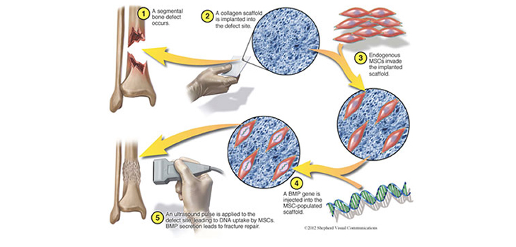
In recent years researchers have tried to grow new bone without using a graft. They first fill gaps in bone with a scaffolding material—often a form of collagen or synthetic grafting material. This material is meant to help the patient’s own bone-forming cells, capillaries and signals to engulf the scaffold use that structure to construct new bone.
For too many patients, particularly the elderly or diabetic or otherwise compromised by such treatments as chemotherapy, those natural healing processes do not always turn into osteocytes, the bone-producing cells. Sometimes they develop into either fat tissue cells or scar tissue.
How to get the patients naturally occurring progenitor cells and other tissues to turn into bone or more bone morphogenetic proteins (BMPs) was the primary research challenge. So investigators decided to use a virus to introduce extra copies of BMP genes into a particular form of progenitor cell which is part of the bone forming lineage in the hope that those cells would produce the proteins long enough to trigger their own differentiation. But this strategy carries risks of malignancy and immune responses.
Then Dan Gazit, D.M.D., Ph.D., a regenerative medicine expert at Cedars-Sinai and his colleagues, packed a wound with the usual collagen matrix and let it sit for a couple of weeks to give progenitor cells time to infiltrate the scaffold. Then they made a solution containing copies of their gene of interest alongside gas-filled micron-sized bubbles encased in a shell of fat molecules. They injected this solution into the fracture sites, and then went over the area with an ultrasound wand. The wand’s ultrasound pulses burst the microbubbles, punching nano-sized holes in the adjacent stem cells, which allowed the genes in the solution to enter.
Gazit’s group used that approach to insert copies of the gene for BMP-6 into pigs that had surgically been given 1-centimeter gaps in a leg bone. After waiting eight weeks, they found that the bone gap was closed and the leg fracture was healed in every one of the treated animals. The procedure was so effective that the fractures healed as well as if bone grafts had been carried out using bone from the same animal, according to their report in Science Translational Medicine.
The results are “just the type of thing we need to move this field forward,” said Johnny Huard, Ph.D., an orthopedics researcher at the University of Texas Health Science Center in Houston.
Brilliant.
React:
Discussion
This is a fascinating development. In my practice we've seen similar outcomes with the revised protocol. The key differentiator seems to be patient selection criteria. Has anyone else noticed the correlation with BMI thresholds?
Great point. I'd push back slightly on the conclusion, the sample size in the cited study is too small to draw population-level inferences. That said, the directional signal is compelling and worth a larger RCT.
We implemented a similar approach last year. Early results are promising but we're still gathering 12-month follow-up data. Happy to share our protocol if anyone is interested.
Join the conversation
Orthopedic professionals are discussing this. Sign in and upgrade to read every comment and add your voice.