Is that bone fully healed? Can the patient get up and walk around safely? Researchers from the Icahn School of Medicine at Mount Sinai in New York, who presented their results at the 2017 meeting of the American Academy of Orthopaedic Surgeons, set out to lessen the subjectivity of bone healing during lengthening.
Bone Healing During IM Lengthening: Clarity!
2 min read Premium comments
Secondary
Ettore Vulcano, M.D, assistant professor, orthopedics, Icahn School of Medicine at Mount Sinai. He commented to OTW, “During lengthening of a bone, the necessity for leg length discrepancies, new bone—called regenerate—is formed. This regenerate will mature over time to become solid bone. However, until this happens, the patient cannot walk to prevent fracturing at this site. To date, the assessment of bone healing has been subjective, based on the surgeon’s experience. The aim of this study was to try to identify an objective means of assessing bone healing, regardless of surgeon experience.”
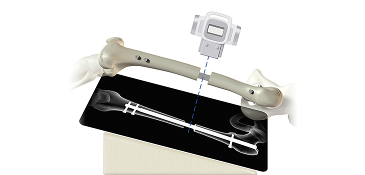
The authors wrote, “…the present study investigated bone regenerate pixel density on a picture archiving and communication systems (PACS) monitor after antegrade femur lengthening using IM [intramedullary] rods. Thirty-two consecutive patients who underwent antegrade femur lengthening using an IM rod at a minimum of 1-year follow up were included in this retrospective study…. Serial, 2-view radiographs of the femur were assessed by a single operator starting at the completion of lengthening (week 0). The pixel density of the lateral, medial, anterior, and posterior cortices was measured in each patient at every postoperative visit. These values were then compared to the adjacent 2 cm of bone just distal to the regenerate. The pixel density ratio (PDR) was calculated, and subsequently correlated to the subjective assessment of bone healing by one of the senior authors.”
Dr. Vulcano told OTW, “We were surprised to find an actual cut-off number that corresponds to good bone healing. Further analysis of the bone density measured with standard X-rays, demonstrated that the bone regenerate continues to mature and to get stronger even one year after surgery. As a matter of fact, it becomes even stronger that the native bone.”
“Having an objective measure of bone healing allows for comparison between patient visits. It allows a surgeon to say whether a bone is healed without looking at the X-rays (as long as a nurse, PA [physician assistant], resident, or colleague measures the bone density value—which requires minimal training). This is great if we consider patients that need to travel long distances to go to their orthopedist’s office. It provides a more accurate and objective assessment of bone healing, translating into safer indications for patients in terms of when they can return to walk fully. In fact, premature weight bearing can lead to catastrophic failure of the rods used to lengthen the bones, whereas excessive non weight bearing can lead to osteoporosis, muscle atrophy, upper extremity pain (walking on crutches), blood clots.”
React:
Discussion
This is a fascinating development. In my practice we've seen similar outcomes with the revised protocol. The key differentiator seems to be patient selection criteria. Has anyone else noticed the correlation with BMI thresholds?
Great point. I'd push back slightly on the conclusion, the sample size in the cited study is too small to draw population-level inferences. That said, the directional signal is compelling and worth a larger RCT.
We implemented a similar approach last year. Early results are promising but we're still gathering 12-month follow-up data. Happy to share our protocol if anyone is interested.
Join the conversation
Orthopedic professionals are discussing this. Sign in and upgrade to read every comment and add your voice.