The Achilles tendon, the strongest tendon in the human body, bears loads as great as ten times the body weight when the subject is jumping. Rainer Burgkart, M.D., senior physician and research director at the Chair of Orthopedics and Sports Orthopedics at the Technical University of Munich (TUM), said of the tendon, “Even though orthopedic doctors treat patients with tendon injuries every day, we still know very little about the precise histological structure at the direct interface between bone and tendon: The biochemical processes, the micromechanics and the microstructure of the tissue have hardly been researched.”
Team of Scientists Reveal Secrets of Achilles Heel
2 min read Premium comments
Secondary
That is no longer the case. Working with a team of doctors, physicists, chemists and engineers at TUM, Burgkart and his colleagues have discovered why the connection between tendon and bone is so strong.
They discovered a tissue layer between the tendons and bones that is composed of extremely thin protein fibers that ensure their remarkable strength. Those fibers are why athletes can take hurdles during a sprint and survive hard landings without damaging the bond between the tendon and ankle bone. In fact, the tendon is more likely to tear than it is to separate from the bone tissue.
The researchers discovered the fine fibers by using a new interdisciplinary research approach. “Up to now, it was thought that the tendons attach directly to the bone. In fact, though, there is a transitional zone. Here the tendon tissue splits into dozens of fine fibers with a characteristic biochemical composition,” explained Prof. Dr. rer. nat. Andreas Bausch, Chair of Cellular Biophysics and director of the interdisciplinary research group. “The thin fibers are firmly anchored to the jagged surface of the bone and are mechanically extremely durable.”
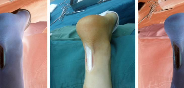
They clamped, in an apparatus, a piece of porcine bone with a tendon attached and aimed a microscope at the boundary layer, along which the tendon and bone grow together. Using multiscale microscope technology, they took dozens of images and compiled them into a single, large image. “In this way, we could make visible the structure of the fine, split fibers,” reports Bausch.
The team used fluorescing antibodies to get specific proteins to light up. From this they learned that the thin fibers have a biochemical composition different from that of the actual tendon. In the third part of their experiment, the researchers moved the tendon back and forth under load and filmed the fibers. The result: Depending on the loading direction, different fibers are active and stabilize the contact.
They augmented the light microscopy investigations by using high-resolution images of an electron microscope. At the Chair of Medical Biophysics the scientists also employed microcomputer tomography to represent the interface region in three dimensions. In addition, researchers at the Chair of Organic Chemistry analyzed the various proteins in the tendons and interface fibers.
“These results allow us, for the first time, to understand the biochemical processes in the contact zone between bones and tendons, which give our locomotor system its extreme strength,” said Bausch.
React:
Discussion
This is a fascinating development. In my practice we've seen similar outcomes with the revised protocol. The key differentiator seems to be patient selection criteria. Has anyone else noticed the correlation with BMI thresholds?
Great point. I'd push back slightly on the conclusion, the sample size in the cited study is too small to draw population-level inferences. That said, the directional signal is compelling and worth a larger RCT.
We implemented a similar approach last year. Early results are promising but we're still gathering 12-month follow-up data. Happy to share our protocol if anyone is interested.
Join the conversation
Orthopedic professionals are discussing this. Sign in and upgrade to read every comment and add your voice.