Royal Philips, based in Amsteram, The Netherlands, is announcing the launch of an “industry-first” augmented-reality surgical navigation technology that is designed to help surgeons perform image-guided open and minimally-invasive spine surgery.
Philips: New Augmented-Reality Navigation Technology
2 min read Premium comments
Secondary
“This unique augmented-reality technology is an example of how we expand our capabilities with innovative solutions in growth areas such as spine, neuro and trauma surgery, ” said Ronald Tabaksblat, business leader Image-Guided Therapy Systems at Philips, in the January 12, 2017 news release.
The results of the first pre-clinical study on the technology have been published in Spine as a result of a collaboration between Philips, Karolinska University, Stockholm, Sweden, and the Cincinnati Children’s Hospital Medical Center. The technology was shown to be significantly better with respect to overall accuracy, compared to pedicle screw placement without the aid of Philips’ augmented-reality surgical navigation technology (85% vs 64%, p<0.05).
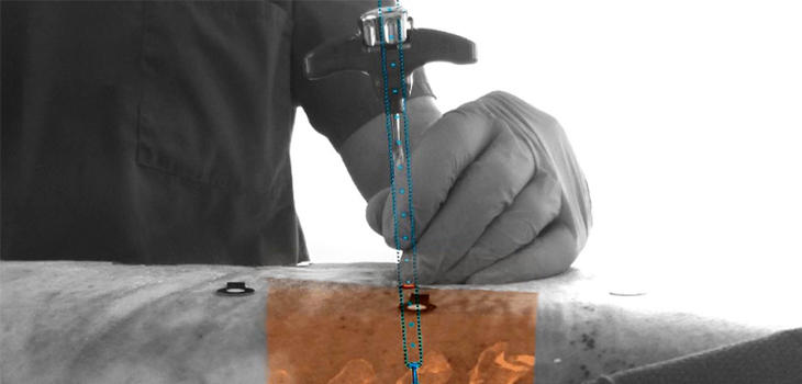
Ronald Tabaksblat told OTW, “The basis is a hybrid room equipped with a Maquet surgical table connected to a motorized ceiling-mounted Carmflat detector system, enabling high-resolution cone-beam CT acquisition. The navigation system, developed by Philips Healthcare, consisted of four high resolution (5 megapixels) optical video cameras mounted in the frame of the flat detector and directed toward the isocenter of the C-arm. Before starting the procedure, 8 to 10 sterile, flat, adhesive circular markers were randomly placed on the skin around the surgical site after skin incision and surgical exposure of the whole thoracic spine.”
“The markers were specifically designed to be tracked by the system. A minimum of five markers had to be in the field of view of the cameras for patient tracking. In order to compensate for any motion, a mesh model generated by interconnecting the markers was used throughout the procedure as a patient tracking model. The flat detector could cover multiple vertebrae in one single rotational scan.”
“All images were displayed on a 58-inch high definition medical monitor. The C-arm automatically rotated into one of the four possible positions where an optical camera was aligned along the planned axis of insertion. The other three optical cameras provided angulated views for the alignment of the devices. The screw path was overlaid on the optical camera views depicting the intended device alignment as well as 2D slices or 3D volume rendering from the acquired CBCT. The optical and intraoperative images were automatically registered and therefore the operator did not interact with the system for registration. During screw insertion, the optical overlay provided real-time feedback to the operator.”
Asked about milestones, he noted, “First was the milestone of technical accuracy of the system: getting the optical cameras into the X-ray flat detector and getting accurate overlays with the 2D and 3D x-ray images.
“The second step was to measure the accuracy of screw placement in a cadaver. This is what we reported in the study in the press release. Third step was the workflow. We wanted it to save time and not take extra time. Together with surgeons from our collaboration clinical sites, we optimized the workflow and user interface. This will continue in 2017 when we go to first-in-human in Sweden.”
React:
Discussion
This is a fascinating development. In my practice we've seen similar outcomes with the revised protocol. The key differentiator seems to be patient selection criteria. Has anyone else noticed the correlation with BMI thresholds?
Great point. I'd push back slightly on the conclusion, the sample size in the cited study is too small to draw population-level inferences. That said, the directional signal is compelling and worth a larger RCT.
We implemented a similar approach last year. Early results are promising but we're still gathering 12-month follow-up data. Happy to share our protocol if anyone is interested.
Join the conversation
Orthopedic professionals are discussing this. Sign in and upgrade to read every comment and add your voice.