Take a crustacean and some live imaging and you have new information of how epidermis cells act during the regrowth of adult limbs after amputation. Researchers from the Institute of Functional Genomics in Lyon (Institut de Génomique Fonctionnelle de Lyon, IGFL), France, have done continuous live imaging of a regenerating leg in the crustacean Parhyale hawaiensis, a close relative of the common sand hopper. Parhyale hawaiensis was introduced in 2014 as a model for regenerative studies in a paper co-authored by Michalis Averof, Ph.D., Director of Research at IGFL and senior author of the current study.
World’s First! Time-Lapse Footage of Limb Regeneration
2 min read Premium comments
Secondary
“Parhyale hawaiensis is well suited for imaging limb regrowth, ” Averof said in the October 25, 2016 news release. “The animals have relatively rapid limb regeneration, requiring as little as one week for young adults to fully regrow their legs. Also, their tiny limbs enable us to image the regeneration process in unprecedented cell-by-cell detail through their entire thickness.”
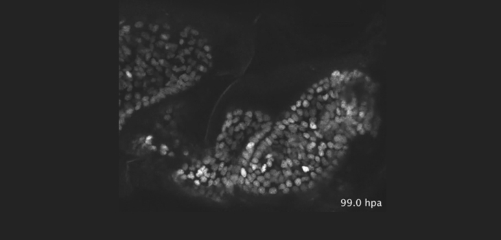
“To record the regrowth of the Parhyale hawaiensis leg, the team used genetically engineered tools to label the limb’s epidermal cells using fluorescent proteins. They then used microscopy to take continuous live recordings over the first days of regrowth, with the fluorescence in the cells allowing them to capture their individual activity.”
“Using this method, we identified a specific sequence of events and cell behaviours that unfold during limb regrowth, ” says first author Frederike Alwes, Ph.D..
“These include wound closure, followed by a quiet period when the epidermal cells migrate slowly towards the site of the wound, which then leads to extensive cell division and movement as the new leg starts to develop its shape. We were surprised to see that there was a sharp transition between the last two stages, which suggests the cells were coordinated by a common signal.”
Alwes explained, “Traditionally, insight into cell behaviour during limb regrowth has been gathered by imaging fixed samples and attempting to fill in the missing pieces, due to the difficulties in tracking cells during regeneration in active adult animals. With the ability to track the movements and behaviour of single cells individually through time, we now have the means to understand the cellular dynamics of the regeneration process, which could not have been reconstructed from fixed material.”
Averof told OTW, “Our main driver was curiosity about how complex tissues and organs regenerate. We know a lot about the mechanisms of development and tissue maintenance in a variety of animals, but very little about organ regeneration. Our experimental organism, a small crustacean, gave us the opportunity to approach this problem by asking how different cells behave and coordinate with each other during leg regeneration.”
“We are excited by the fact that we can image the whole process of leg regeneration continuously. It means that we will be able to have a complete description of how each cell behaves and what its descendants contribute during regeneration.”
React:
Discussion
This is a fascinating development. In my practice we've seen similar outcomes with the revised protocol. The key differentiator seems to be patient selection criteria. Has anyone else noticed the correlation with BMI thresholds?
Great point. I'd push back slightly on the conclusion, the sample size in the cited study is too small to draw population-level inferences. That said, the directional signal is compelling and worth a larger RCT.
We implemented a similar approach last year. Early results are promising but we're still gathering 12-month follow-up data. Happy to share our protocol if anyone is interested.
Join the conversation
Orthopedic professionals are discussing this. Sign in and upgrade to read every comment and add your voice.