Chemists from Trinity College in Dublin, Ireland, and their collaborators with RCSI (Royal College of Surgeons of Ireland) are celebrating their discovery of new scanning techniques that produce extremely high resolution images of cracks in bones—even cracks at a micro level.
Gold Nanoagents in X-Rays Reveal Microcracks in Bones
2 min read Premium comments
Secondary
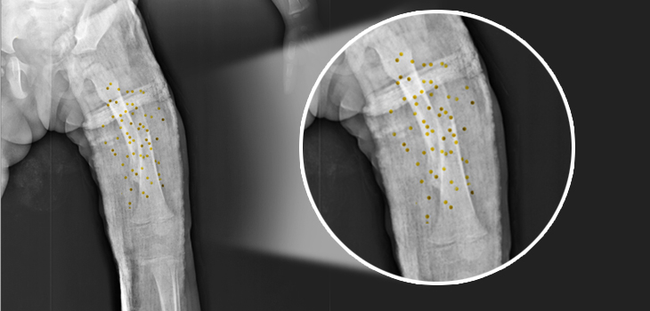
The chemists attach luminescent compounds to tiny gold structures to form biologically safe ‘nanoagents’ that are attracted to calcium-rich surfaces which appear when bones crack—even at a micro level. These nanoagents highlight the cracks formed in bones, allowing researchers to produce a complete 3D image of a damaged region.
The technique, researchers believe, can be used to diagnose bone strength and provide a detailed blueprint of the extent and precise positioning of weakness or injury. This should help prevent the use of bone implants in many cases, and act as an early-warning system for people at risk of degenerative bone diseases, such as osteoporosis.
The research, led by the Trinity team of Professor of Chemistry Thorri Gunnlaugsson, Ph.D., and Postdoctoral Researcher Esther Surender, Ph.D., has been published in the journal Chem, a sister journal to Cell. Gunnlaugsson said, “We have demonstrated that we can achieve a three-dimensional map of bone damage, showing the microcracks, using non-invasive luminescence imaging. The nanoagent we have developed allows us to visualize the nature and the extent of the damage in a manner that wasn’t previously possible. This is a major step forward in our endeavour to develop targeted contrast agents for bone diagnostics for use in clinical applications.”
RCSI Professor of Anatomy Clive Lee, Ph.D., M.D. noted that everyday activity loads bones and causes microcracks to develop. These are normally repaired by the body’s remodeling process, but, when microcracks develop faster, they can exceed the repair rate and so accumulate and weaken bones. He says that this occurs in “athletes and leads to stress fractures. In elderly people with osteoporosis, microcracks accumulate because repair is compromised. This leads, he notes, to fragility fractures, most commonly in the hip, wrist and spine.” Current X-ray techniques can tell us about the quantity of bone present but they do not give much information about bone quality, ” he said.
By using their new nanoagent to label microcracks and detect them with magnetic resonance imaging, the researchers hope to measure both bone quantity and quality and identify those at greatest risk of fracture. “Diagnosing weak bones before they break should reduce the need for operations and implants—prevention is better than cure, ” said Lee.
Dr. Surender believes that the nanoagents have great potential for clinical application. She says that by using gold nanoparticles, they were able to lower the overall concentration of the agent that would have to be administered within the body, which is ideal from a clinical perspective. By using what is called ‘two-photon excitation’ they were able to image bone structure using long wavelength excitation, which, they noted, is not harmful or damaging to biological tissues.
React:
Discussion
This is a fascinating development. In my practice we've seen similar outcomes with the revised protocol. The key differentiator seems to be patient selection criteria. Has anyone else noticed the correlation with BMI thresholds?
Great point. I'd push back slightly on the conclusion, the sample size in the cited study is too small to draw population-level inferences. That said, the directional signal is compelling and worth a larger RCT.
We implemented a similar approach last year. Early results are promising but we're still gathering 12-month follow-up data. Happy to share our protocol if anyone is interested.
Join the conversation
Orthopedic professionals are discussing this. Sign in and upgrade to read every comment and add your voice.