Want to turn your patients’ CT and MRI scans into 3D simulations? Zimmer Biomet Holdings, Inc. has a new app for that after acquiring a Dutch technology company which developed an app that converts medical scans into 3D motion simulations of a patients’ anatomy.
Zimmer Biomet Acquires 3D Simulation App
2 min read Premium comments
Secondary
On September 15, 2016, the company announced it acquired Clinical Graphics, B.V. for an undisclosed amount of money. The announcement stated the company plans to integrate the new imaging platform to enhance its hip preservation portfolio.
Clinical Graphics, started in 2010 as a spin-off from Delft University of Technology, is a pioneer in the field of 3D interactive range-of-motion simulation reports for common hip conditions such as femoroacetabular impingement (FAI) and dysplasia. Physicians use the technology to characterize the physiology of a patient’s pain and direct treatment approaches.
The simulation service, called Move Forward, requires no specialized software, hardware or training.
Here’s How It Works
Physicians order a free “On-the-GO! First Aid Kit.” The kit contains a USB flash drive with an introductory video and the GO! upload application.
After uploading the app, users create an account and start uploading CT or MRI scans. The app helps users to find their DICOM (Digital Imaging and Communications in Medicine) files, compresses and anonymizes the files before uploading them. No information is submitted that will allow identification of a patient. GO! scrambles the files with a security key. The image data is completely unreadable until it reaches Clinical Graphics’ processing computers.
Once the scan is processed the company emails users a download link to their 3D motion simulation report.
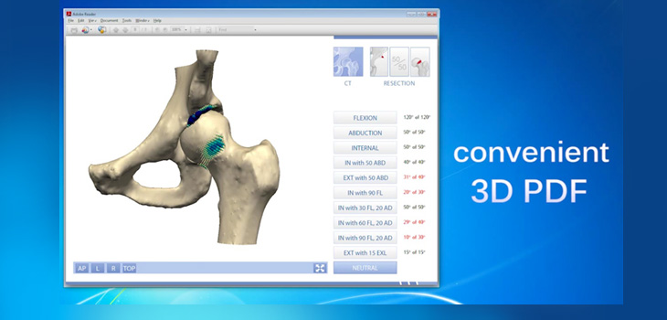
Here’s one example from the Clinical Graphics website for a femoroacetabular impingement report:
The FAI reports contain 10 motion tests, ranging from basic flexion to complex combined motion patterns. Users can find the exact point of contact and find out where bone-on-bone impingement occurs. Reports include radiographic measurements and simulated resection plans.
The Clinical Graphics site states that traditional imaging modalities such as CT or MRI sometimes fail to depict the bone shape variations that may lead to hip impingement. The complex relationship between the acetabular cup and the femoral head and how these interact during motion is difficult to grasp without an actual visual representation. The company’s app provides an interactive 3D view of the hip joint in motion.
On request, the company can also add custom 3D motion tests. For instance, gymnastics require different motion patterns than hurdle runners. In addition to these advanced calculations, the reports include radiographic parameters such as acetabular version, joint coverage, alpha angles and LCE angles.
Dan Williams, Zimmer Biomet’s joint reconstruction group president, said, “3D imaging represents the next generation of treating joint pain, and we’re excited to team up with Clinical Graphics and integrate our technologies to further enhance the clinical utility of our market leading hip portfolio.”
React:
Discussion
This is a fascinating development. In my practice we've seen similar outcomes with the revised protocol. The key differentiator seems to be patient selection criteria. Has anyone else noticed the correlation with BMI thresholds?
Great point. I'd push back slightly on the conclusion, the sample size in the cited study is too small to draw population-level inferences. That said, the directional signal is compelling and worth a larger RCT.
We implemented a similar approach last year. Early results are promising but we're still gathering 12-month follow-up data. Happy to share our protocol if anyone is interested.
Join the conversation
Orthopedic professionals are discussing this. Sign in and upgrade to read every comment and add your voice.