You can remain standing, it can handle both knees at once…what’s not to like? Pennsylvania-based CurveBeam is presenting its new orthopedic computed tomography (CT) scanner aimed at the knees and lower extremities.
CurveBeam Announces Development of Extremity CT System for Knee
2 min read Premium comments
Secondary
Neil Segal, M.D., a specialist in rehabilitation medicine, has been overseeing research efforts using a LineUP prototype, first at the University of Iowa and currently at the University of Kansas. Dr. Segal said in the September 15, 2016 news release, “Difficulty in reproducing the same view of the joint over time impairs ability to detect joint disease, and the 2D nature of radiographs makes these images of overlapping bony anatomy very insensitive for detecting abnormalities until there is advanced joint damage.”
As indicated in the news release, the LineUP will provide isotropic, three-dimensional volumes of the anatomy with a high-resolution output of between 0.2 mm and 0.3 mm slices. The LineUP will be the only cone beam CT system that can provide bilateral, weight bearing scans.
“A study led by Dr. Segal focused on osteophytes, one structure linked to pain in people with knee osteoarthritis. Knees of community-dwelling adults with knee OA were imaged with MRI (reference), fixed-flexion radiographs, and weight bearing CT. The sensitivity and accuracy for detecting osteophytes and subchondral cysts were higher with weight bearing CT imaging in comparison to fixed-flexion radiographs. The study was published in the August 2016 issue of the Journal of Orthopedic Research.”
“Clinically, this is a highly meaningful improvement, ” Dr. Segal said. “It suggests that weight-bearing CT could replace radiographs as the recommended means of assessing knee OA. This advancement is even more significant given that it was made without significantly increasing the radiation dose (0.01 mSv [millisievert] for SCT vs. 0.005–0.102 mSv for a series of knee radiographs).”
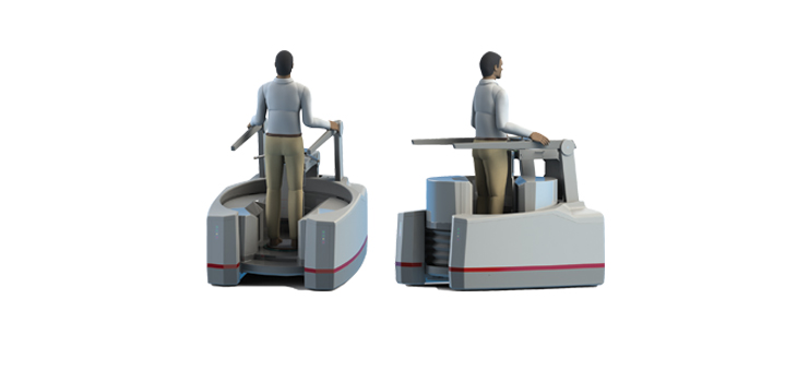
Vinti Singh of CurveBeam Sales & Marketing, told OTW, “The LineUP can do a CT scan of a patient while he or she is standing. This is not possible in a conventional medical CT scanner. The device is also compact enough that it can fit in an orthopedic clinic. The LineUP is the only extremity CT scanner that can scan both knees at once. Other extremity CT scanners can only scan one knee at a time, which means the patient is not standing naturally, but rather balanced on one leg. The benefit of standing CT scans are to see the bones and joint in weight bearing position, which is not achievable unless a patient is standing balanced on both feet.”
“We want orthopedic surgeons to know it is possible to get a standing, weight bearing scan of the knees, and that they can take these scans in their own office and bill for them.”
React:
Discussion
This is a fascinating development. In my practice we've seen similar outcomes with the revised protocol. The key differentiator seems to be patient selection criteria. Has anyone else noticed the correlation with BMI thresholds?
Great point. I'd push back slightly on the conclusion, the sample size in the cited study is too small to draw population-level inferences. That said, the directional signal is compelling and worth a larger RCT.
We implemented a similar approach last year. Early results are promising but we're still gathering 12-month follow-up data. Happy to share our protocol if anyone is interested.
Join the conversation
Orthopedic professionals are discussing this. Sign in and upgrade to read every comment and add your voice.