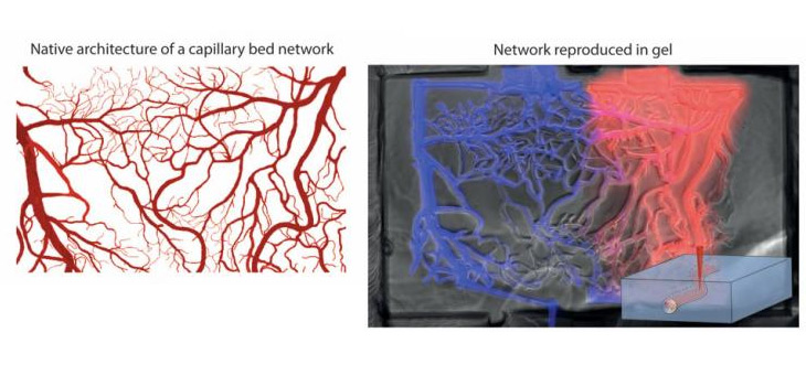
Researchers from Switzerland have found a way to expand our ability to work with cell cultures. Matthias Lütolf and his Ph.D. student Nathalie Brandenberg, working at the Ecole Polytechnique Federale de Lausanne, have found a way to use lasers to influence cell functioning. As indicated in the June 23, 2016 news release, “So far, most cell culture approaches are limited to two-dimensional environments (e.g., a Petri dish or a chip), but that neither matches real biology nor helps us sculpt tissues and organs. Two EPFL scientists have now developed a new method that uses lasers to carve out paths inside biocompatible gels to locally influence cell function and promote tissue formation. The work is published in Advanced Materials.”
Lasers to Reduce Risk of Necrosis?
2 min read Premium comments
Secondary
“The team has developed a method that uses a laser to cut three-dimensional pathways and networks for cells inside a hydrogel scaffold that matches their natural environment. The method combines lasers with microfluidics—the science of controlling fluids in micrometer-sized spaces. The scientists used focalized short-pulsed lasers, which can generate enough power to create tiny tunnels in different gels already used in cell biology and tissue engineering. The laser can be applied before or even during 3D cell culture, meaning that the cells can be controlled in ‘real time’ to match their natural growth.”
Dr. Lütolf told OTW, “We think that this technique could also be used to address the issue of a lacking vasculature (i.e., limited diffusion of nutrients and growth factors) in larger tissue engineered constructs such as bone. As you may know, cells inside scaffolds that are larger than a millimetre or so really suffer, often resulting in necrosis of the construct. Using our laser-based approach, one could generate an artificial blood vessel network (basically an open network of channels) which would result in enhanced diffusion of nutrients, reducing the risk of necrosis quite dramatically, we think.”
“What we are most excited about right now is to use this new tool to generate tissue and organ models in a cell culture dish. We are using lasers to influence and enhance the in vitro development of organ-like structures called ‘organoids’. Organoids have a huge potential for applications in pharmaceutical drug development and toxicology. Current organoid systems are somewhat limited because they grow very uncontrollably and are thus poorly reproducible. Our approach might help to solve this problem.”
React:
Discussion
This is a fascinating development. In my practice we've seen similar outcomes with the revised protocol. The key differentiator seems to be patient selection criteria. Has anyone else noticed the correlation with BMI thresholds?
Great point. I'd push back slightly on the conclusion, the sample size in the cited study is too small to draw population-level inferences. That said, the directional signal is compelling and worth a larger RCT.
We implemented a similar approach last year. Early results are promising but we're still gathering 12-month follow-up data. Happy to share our protocol if anyone is interested.
Join the conversation
Orthopedic professionals are discussing this. Sign in and upgrade to read every comment and add your voice.