A new technique developed by Gordana Vunjak-Novakovic, Ph.D., the Mikati Foundation Professor of Biomedical Engineering at Columbia Engineering and professor of medical sciences (in Medicine) at Columbia University, repairs large bone defects in the head and face by using lab-grown living bone, tailored to the patient and the defect being treated. According to the June 15, 2016 news release, “This is the first time researchers have grown living bone that precisely replicates the original anatomical structure, using autologous stem cells derived from a small sample of the recipient’s fat.”
Growing Bone Replicates Anatomical Structure
2 min read Premium comments
Secondary
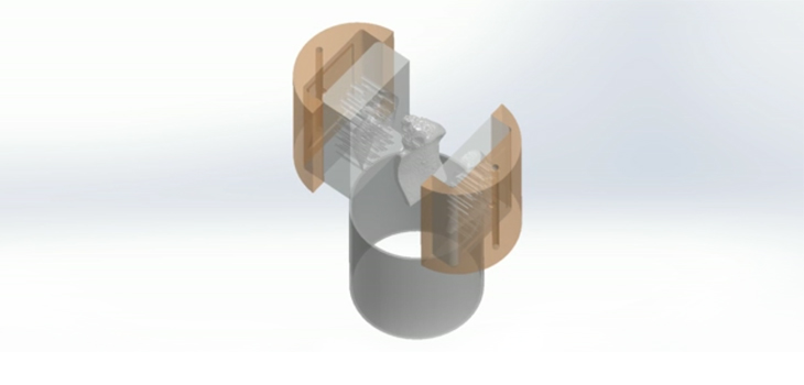
Dr. Vunjak-Novakovic told OTW, “The technology is to repair large bone defects in head and face by using lab-grown living bone, tailored to the patient and defect being treated. The bone is formed by the recipient’s own cells (obtained from a small fat aspirate), within a scaffold made from bone matrix, in a perfused bioreactor. The scaffold and bioreactor chamber are fabricated based on the images of the defect, to provide a perfect anatomical fit.
“The particular scaffold we use allows bone formation without the use of growth factors, and at the same time provides mechanical competence under loading, both of which are unique advantages for clinical application. We compared implantations of engineered bone and a scaffold, both tailored to anatomical shapes, and showed major advantages of using living bone. Our bones maintain their health by a constant remodeling—resorption of some of the old bone, and its replacement by the new bone. Interestingly, while both the bone and scaffold grafts were processed by the body, only the bone grafts had biological ability to drive synthesis of new bone.
“Finally, and unexpectedly, the lab-grown bone, when implanted, was gradually replaced by new bone formed by the body, this serving as a template for bone formation rather than as a definitive implant. This feature is what makes this implant ‘your own bone’ that will become an integral part of the native bone.”
Regarding future research, she noted, “Because bone regenerates more readily than many other tissues, we are much closer to clinical application of this technology than most other applications, which is another reason that we are diligently working towards clinical trials. To make this happen, I founded a company epiBone with the students who participated in these studies. In parallel, we are using the bone-cartilage grafts as models of high biological fidelity in studies of skeletal regeneration and disease.”
React:
Discussion
This is a fascinating development. In my practice we've seen similar outcomes with the revised protocol. The key differentiator seems to be patient selection criteria. Has anyone else noticed the correlation with BMI thresholds?
Great point. I'd push back slightly on the conclusion, the sample size in the cited study is too small to draw population-level inferences. That said, the directional signal is compelling and worth a larger RCT.
We implemented a similar approach last year. Early results are promising but we're still gathering 12-month follow-up data. Happy to share our protocol if anyone is interested.
Join the conversation
Orthopedic professionals are discussing this. Sign in and upgrade to read every comment and add your voice.