A team from The Johns Hopkins University has found a way to use manmade plastic to create a framework for filling in missing bone. According to the May 4, 2016 news release, it involves mixing at least 3% pulverized natural bone with the plastic and creating the necessary shape with a 3D printer.
Recipe For Missing Bone: Found!
2 min read Premium comments
Secondary
Warren Grayson, Ph.D. associate professor of biomedical engineering at the Johns Hopkins University School of Medicine was the report’s senior author. He and his team used polycaprolactone, or PCL, a biodegradable polyester used in making polyurethane that has been approved by the FDA for other clinical uses.
To strengthen the PCL, they mixed it with increasing amounts of bone powder, made by “pulverizing the porous bone inside cow knees after stripping it of cells.”
“After three weeks, cells grown on 70% bone powder scaffolds showed gene activity hundreds of times higher in three genes indicative of bone formation, compared to cells grown on pure PCL scaffolds. Cells on 30% bone powder scaffolds showed large but less impressive increases in the same genes.”
“After the scientists added the key ingredient beta-glycerophosphate to the cells’ broth to enable their enzymes to deposit calcium, the primary mineral in bone, the cells on 30 percent scaffolds produced about 30 percent more calcium per cell, while those on 70 percent scaffolds produced more than twice as much calcium per cell, compared to those on pure PCL scaffolds.”
“Finally, the team tested their scaffolds in mice with relatively large holes in their skull bones made experimentally. Without intervention, the bone wounds were too large to heal. Mice that got scaffold implants laden with stem cells had new bone growth within the hole over the 12 weeks of the experiment. And CT scans showed that at least 50% more bone grew in scaffolds containing 30 or 70% bone powder, compared to those with pure PCL.”
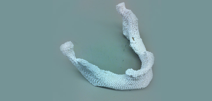
Dr. Grayson told OTW, “The most interesting part of this research was finding out that we could print these scaffolds that contain up to 70% bone by mass! That means the vast majority of these porous scaffolds were made up of native material (which is used clinically), that could be printed in any anatomical shape.”
“Based on the considerable advances that are being made in the biomaterials field, the not-too-distant future may hold the potential for printing out patient-specific replacements grafts made almost entirely of native bone scaffolding. This will overcome any limitations in supply of the patient’s own bone or allografts from bone banks.”
React:
Discussion
This is a fascinating development. In my practice we've seen similar outcomes with the revised protocol. The key differentiator seems to be patient selection criteria. Has anyone else noticed the correlation with BMI thresholds?
Great point. I'd push back slightly on the conclusion, the sample size in the cited study is too small to draw population-level inferences. That said, the directional signal is compelling and worth a larger RCT.
We implemented a similar approach last year. Early results are promising but we're still gathering 12-month follow-up data. Happy to share our protocol if anyone is interested.
Join the conversation
Orthopedic professionals are discussing this. Sign in and upgrade to read every comment and add your voice.