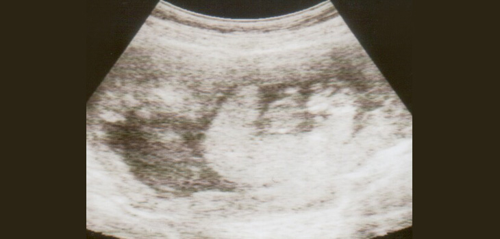
In a long-ranging research effort, years ago, scientists from the UK used data from the Avon Longitudinal Study of Parents and Children (ALSPAC) and found that greater placental size at birth was associated with larger bones at each age in childhood.
Larger Placentae at Birth Associated With Larger Childhood Bones
2 min read Premium comments
Secondary
Researchers at the Medical Research Council Lifecourse Epidemiology Unit, University of Southampton, teamed up with colleagues from the University of Bristol, studied 518 children who underwent bone scans at 9, 15 and 17 years old.
According to the May 3, 2016 news release, “The study, published in the Journal of Bone and Mineral Research, found that the relationship between the placenta and offspring bone remained robust even after adjusting for factors such as the child’s height and weight and pubertal status.”
“The Southampton team believe this latest research offers new insights into earlier observations linking maternal factors in pregnancy with offspring bone health. Larger bones in early life are likely to lead to larger, stronger bones in older adulthood, which reduces the risk of osteoporosis and broken bones in later life. However, more research is needed to understand the more detailed mechanisms underlying associations between placenta size/function and offspring bone mass, the team says.”
Professor Nicholas Harvey, Professor of Rheumatology and Clinical Epidemiology, who led the research in Southampton, commented: “Whilst there are many factors which are likely to influence placental size and function, and importantly, we don’t know as yet whether a larger placenta actually causes the greater offspring bone mass, these findings really help us to understand the possible mechanisms whereby factors such as maternal diet, smoking, physical activity and vitamin D status may influence offspring bone development.”
“This work builds on our previous findings from the Southampton Women’s Survey, and demonstrates that positive associations between placental size and offspring bone size are maintained even through puberty.” Professor Cyrus Cooper, Professor of Rheumatology and Director of the MRC Lifecourse Epidemiology Unit, added, “This work forms part of a larger programme of research seeking to develop interventions in early life aimed at optimising bone development and reducing the risk of osteoporotic fracture in older age. Confirmation of our earlier Southampton findings in the Bristol cohort is testament to the close working between Southampton and Bristol collaborators, and demonstrates the clear benefit to UK science from such cross-cohort investigations.”
Lead researcher Nick Maguire told OTW, “Interestingly, although greater placental size was associated with greater bone size, it was associated with lower volumetric bone density during puberty. This is consistent with the known increased risk of childhood fracture during the early pubertal years, during a phase when bones are growing rapidly in size, but mineralisation lags behind. Fortunately, bone density catches up with bone size in early adult hood, so overall the long-term association will be between greater placental size and stronger adult bones.”
“Overall, this paper emphasises the importance, to all those involved in the care of patients with osteoporosis and fractures, that the risk of osteoporosis begins to be accrued very early in life, even in utero, and thus the prevention of fragility fractures should be considered over the whole lifecourse.”
React:
Discussion
This is a fascinating development. In my practice we've seen similar outcomes with the revised protocol. The key differentiator seems to be patient selection criteria. Has anyone else noticed the correlation with BMI thresholds?
Great point. I'd push back slightly on the conclusion, the sample size in the cited study is too small to draw population-level inferences. That said, the directional signal is compelling and worth a larger RCT.
We implemented a similar approach last year. Early results are promising but we're still gathering 12-month follow-up data. Happy to share our protocol if anyone is interested.
Join the conversation
Orthopedic professionals are discussing this. Sign in and upgrade to read every comment and add your voice.