Mind-Controlled Arm Allows Finger Wiggling
Mind-Controlled Arm Allows Finger Wiggling // Non-Opioid Care Improves Outcomes for Those on Opioids for Pain // and More!
5 min read Premium comments
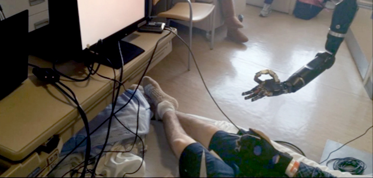
It’s a first! Using 128 electrode sensors—all placed on a single rectangular sheet of film the size of a credit card—researchers at Johns Hopkins University have made inroads with efforts to give prosthetic hands more flexibility. Actually, the team has made it possible for a mind-controlled arm to wiggle fingers individually and independently of each other.
The proof-of-concept feat was described online this week in the Journal of Neural Engineering.
Nathan Crone, M.D., professor of neurology at the Johns Hopkins University School of Medicine, told OTW, “While there have been many people who have worked on a brain/machine interface, we have taken a different approach. Traditional efforts involved taking recordings from as many single neurons as possible (usually up to about 200) from a small area in the motor region of the brain. One downside is that patients have a big learning curve; the other is that you can’t know what the individual neurons are doing. In addition, it’s not easy to maintain recordings of those neurons over time.”
“Our work involved mapping and tracking the specific parts of the subject’s brain that is responsible for moving each finger. While this work could eventually apply to orthopedic patients, we are now working with epilepsy patients. As part of epilepsy surgery the physician implants electrodes on the surface of the brain in order to map where the seizures are coming from and where the critical cortical functions are located so that those can be avoided when treating seizures. If the electrodes happen to be over the motor or sensory cortex for the arm then we can ask the patient to contribute time while they are waiting for a seizure to be recorded. This helps us know whether they can achieve control over the prosthetic limb. This particular patient was having seizures that were emanating from the arm area of the brain, so we had high definition electrodes over that area and could thus map the arm and individual fingers of the hand. That is what allowed the patient to achieve control over the fingers of the prosthetic limb in a way that couldn’t be done before. Because we had mapped out the finger representations in the brain, the patient could control the fingers more intuitively without any training.”
“This initiative falls under a program called Revolutionizing Prosthetics, an ongoing project of the Defense Advanced Research Projects Agency (DARPA). The robotic limb that we used was designed and manufactured at the Applied Physics Lab at Hopkins. This is only the beginning.”
Non-Opioid Care Improves Outcomes for Those on Opioids for Pain
New research from Vanderbilt University Medical Center, Massachusetts General Hospital and Brigham and Women’s Hospital has found that for patients taking opioids for pain, improvements in their functioning are further improved by using multiple non-opioid treatments during hospitalization. Specifically, the readmission rate, length of stay and opioid dose dropped as the use of the non-opioid treatment modalities grew.
David A. Edwards, M.D., Ph.D., clinical chief of Pain at Vanderbilt University Medical Center, was the study’s lead author. He told OTW, “This work originated after Massachusetts General Hospital identified that a subset of patients were dissatisfied with their care, and this group were also opioid tolerant. Upon further investigation this subset of patients were also found to be high utilizers of hospital resources, and also to experience a higher rate of complications during their hospital stay. It was decided by hospital leadership to design a system to identify patients at risk and to minimize their suffering and improve their outcomes.”
“Surgical services understand that patients on a high dose of opioids often are difficult to treat following surgery. These patients stay longer in the hospital and return to the hospital at much higher rates than other patients. To decrease the length of stay and to decrease the readmission rate, all services that interact with the patients should implement evidence from their field shown to impact outcomes for this cohort. By bringing together the evidence into patient care pathways, we hypothesize that patients on high dose opioids will benefit, and hospital systems will become more efficient.”
Asked how the RCT will proceed, Dr. Edwards noted, “The trial is now recruiting patients under the direction of Dr. Padma Gulur at the University of California Irvine and associated California hospitals. Vanderbilt University and Massachusetts General Hospital are in pre-recruitment phase. We anticipate completion of the trial and publication of results over the next two years.”
“Practitioners should know which of their patients meet the definition of opioid tolerant. Then they should know what avenues for pain control are available in their institutions. Non-opioid options for pain control should become familiar to all members of the team and used especially for this higher risk group. For example, in order to prevent problems from occurring with this group of patients, one may need to consider extra education of the patients either by educated team members, including pharmacy, multimodal analgesia, articular analgesia or regional anesthesia, monitoring the dose escalation rate of opioids, and creating a robust discharge system where the patients know who to call or where to go if they have problems.”
New—Biologic—Method of Joint Resurfacing
Using a canine model, researchers at the University of Missouri School of Medicine have found evidence that a new biologic method of resurfacing a joint could result in improved outcomes. The method was developed at the Missouri Orthopaedic Institute. Brett Crist, M.D., an associate professor of orthopaedic surgery at the MU School of Medicine, lead author of the study.
Dr. Crist told OTW, “Instead of using several small, cylinder-shaped bone or cartilage plugs, we used larger, size-matched grafts to cover the area in need of repair. In our dog model, we found that the animals implanted with traditional small grafts showed significant loss in range of motion and joint integrity after only eight weeks. In contrast, the dogs implanted with larger, bevel-shaped grafts maintained joint viability and structural integrity throughout the six-month study period.”
“We think that the success came from reducing the number of seams for a smoother functioning joint; it was also helpful that we beveled the edges as this created a better fitting repair that was less prone to cell death during implantation.”
“We thought the large grafts would do better, but the surprise was that the small grafts didn’t make it to the six month postoperative examination. We thought they would do better than that. It was impressive that none of them made it past the eight week assessment. When doing osteoarticular allografts, it appears that large grafts actually do better and that this is a viable option for people.”
As about future research, Dr. Crist commented to OTW, “The goal is to obtain prospective data on human patients showing the effectiveness of these large osteoarticular femoral head grafts. We want to evaluate their functional recovery over 5 years.”
React:
Discussion
This is a fascinating development. In my practice we've seen similar outcomes with the revised protocol. The key differentiator seems to be patient selection criteria. Has anyone else noticed the correlation with BMI thresholds?
Great point. I'd push back slightly on the conclusion, the sample size in the cited study is too small to draw population-level inferences. That said, the directional signal is compelling and worth a larger RCT.
We implemented a similar approach last year. Early results are promising but we're still gathering 12-month follow-up data. Happy to share our protocol if anyone is interested.
Join the conversation
Orthopedic professionals are discussing this. Sign in and upgrade to read every comment and add your voice.