A group of researchers has taken bio-printing to a new level. They have found a way to 3D bioprint thick vascularized tissue constructs composed of human stem cells, extracellular matrix, and circulatory channels lined with endothelial blood vessel cells. The resulting network of vasculature can channel fluids, nutrients and cell growth factors, for the first time, throughout tissues.
3D Bioprinting Creates Blood Vessels
2 min read Premium comments
Secondary
As Jennifer A. Lewis, Sc.D., senior author on the study and the Hansörg Wyss Professor of Biologically Inspired Engineering at Harvard’s John A. Paulson School for Engineering and Applied Sciences (SEAS), explained, “This latest work extends the capabilities of our multi-material bioprinting platform to thick human tissues, bringing us one step closer to creating architectures for tissue repair and regeneration.”
Until now, scaling up human tissues built of cell types has been limited by an inability to embed life-sustaining vascular networks. Lewis and her team solved this problem by increasing the tissue thickness nearly tenfold. They combined vascular plumbing with living cells and an extracellular matrix which enabled the structures to function as living tissues. They showed that their 3D bioprinted tissues could function as living tissue architectures for upwards of six weeks.
“This research will help to establish the fundamental scientific understanding required for bioprinting of vascularized living tissues, ” said Zhijian Pei, Ph.D., National Science Foundation Program Director for the Directorate for Engineering Division of Civil, Mechanical and Manufacturing Innovation, which funded the project.
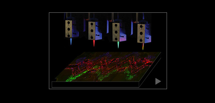
According to Medical Press, Lewis uses a printed silicone mold to house and plumb the printed tissue structure. “Inside this mold, a grid of vascular channels is printed first, over which ink containing living stem cells is then printed. The inks are self-supporting and strong enough to hold shape as the structure’s size increases with each layer of deposition. At intersections meeting within the foundational vascular grid, vertical vascular pillars are printed, which interconnect a network of microvessels. After printing, a liquid composed of fibroblasts and extracellular matrix fills in the open regions around the 3D printed tissue, cross linking the entire structure.”
The resulting structure has a quantity of blood vessels, and can be immediately perfused with nutrients to ensure survival of the cells.
Team members for this breakthrough research came from the Wyss Institute for Biologically Inspired Engineering at Harvard University and the Harvard John A. Paulson School for Engineering and Applied Sciences. The full story is reported in the journal Proceedings of the National Academy of Sciences.
React:
Discussion
This is a fascinating development. In my practice we've seen similar outcomes with the revised protocol. The key differentiator seems to be patient selection criteria. Has anyone else noticed the correlation with BMI thresholds?
Great point. I'd push back slightly on the conclusion, the sample size in the cited study is too small to draw population-level inferences. That said, the directional signal is compelling and worth a larger RCT.
We implemented a similar approach last year. Early results are promising but we're still gathering 12-month follow-up data. Happy to share our protocol if anyone is interested.
Join the conversation
Orthopedic professionals are discussing this. Sign in and upgrade to read every comment and add your voice.