Armed with funding from the Armed Forces Institute of Regenerative Medicine, researchers at Wake Forest Baptist Medical Center have designed a special 3D printer and proven that it is possible to print living tissue structures to replace injured or diseased tissue in patients.
Custom 3D Printer Used to Print Living Tissue
2 min read Premium comments
Secondary
As indicated in the February 15, 2016 news release, “Reporting in Nature Biotechnology, the scientists said they printed ear, bone and muscle structures. When implanted in animals, the structures matured into functional tissue and developed a system of blood vessels. Most importantly, these early results indicate that the structures have the right size, strength and function for use in humans.”
“This novel tissue and organ printer is an important advance in our quest to make replacement tissue for patients, ” said Anthony Atala, M.D., director of the Wake Forest Institute for Regenerative Medicine (WFIRM) and senior author on the study, in the news release. “It can fabricate stable, human-scale tissue of any shape. With further development, this technology could potentially be used to print living tissue and organ structures for surgical implantation.”
Dr. Atala told OTW, “Our research indicates the feasibility of printing bone, muscle and cartilage for patients. The results of this study bring us closer to the reality of using 3D printing to repair defects using the patient’s own engineered tissue. It is often frustrating for physicians to have patients receive a plastic or metal part during surgery knowing that the best replacement would have been the patient’s own tissue.”
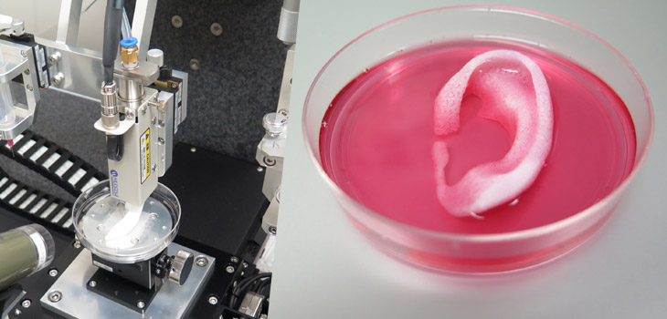
According to the news release, “The Integrated Tissue and Organ Printing System (ITOP), developed over a 10-year period by scientists at the Institute for Regenerative Medicine, overcomes these challenges. The system deposits both bio-degradable, plastic-like materials to form the tissue “shape” and water-based gels that contain the cells. In addition, a strong, temporary outer structure is formed. The printing process does not harm the cells.”
“A major challenge of tissue engineering is ensuring that implanted structures live long enough to integrate with the body. The Wake Forest Baptist scientists addressed this in two ways. They optimized the water-based “ink” that holds the cells so that it promotes cell health and growth and they printed a lattice of micro-channels throughout the structures. These channels allow nutrients and oxygen from the body to diffuse into the structures and keep them live while they develop a system of blood vessels.”
“It has been previously shown that tissue structures without ready-made blood vessels must be smaller than 200 microns (0.007 inches) for cells to survive. In these studies, a baby-sized ear structure (1.5 inches) survived and showed signs of vascularization at one and two months after implantation.”
“And, to show that construction of a human-sized bone structure, jaw bone fragments were printed using human stem cells. The fragments were the size and shape needed for facial reconstruction in humans. To study the maturation of bioprinted bone in the body, printed segments of skull bone were implanted in rats. After five months, the bioprinted structures had formed vascularized bone tissue.”
React:
Discussion
This is a fascinating development. In my practice we've seen similar outcomes with the revised protocol. The key differentiator seems to be patient selection criteria. Has anyone else noticed the correlation with BMI thresholds?
Great point. I'd push back slightly on the conclusion, the sample size in the cited study is too small to draw population-level inferences. That said, the directional signal is compelling and worth a larger RCT.
We implemented a similar approach last year. Early results are promising but we're still gathering 12-month follow-up data. Happy to share our protocol if anyone is interested.
Join the conversation
Orthopedic professionals are discussing this. Sign in and upgrade to read every comment and add your voice.