A huge new study from the University of Southampton in the UK has found that lesions, which are visible on MRI scans, could help identify individuals who are more likely to have more rapidly progressing osteoarthritis. The work was conducted by the university’s Medical Research Council Lifecourse Epidemiology Unit and has been published in The Journal of Rheumatology.
Study: Bone Marrow Lesions Identify Rapid Osteoarthritis
2 min read Premium comments
Secondary
According to the January 15, 2016 news release, “The SEKOIA study, an international osteoarthritis disease-modifying trial, carried out MRI scanning on the knees of 176 men and women over 50 years old. They were then followed up for an average of three years with repeated knee X-rays. Individuals with abnormalities on their MRI scans at the first appointment were compared to those without to examine the effect on disease progression.”
“Individuals with bone marrow lesions (BMLs) on their MRI scan were found to have osteoarthritis that progressed more rapidly than those that did not. On average, the space within the joint is lost at a rate of 0.15mm per year however the Southampton study shows that, overall, individuals with BMLs had a loss rate that was 0.10mm per year faster than those without BMLs. This may lead to earlier need for joint replacement or other intervention.”
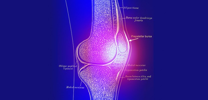
“BMLs show up on MRI as regions of bone beneath the cartilage with ill-defined high signal and represent areas of bone marrow edema, fibrosis, and necrosis. The Southampton researchers believe that therapies to target these abnormalities may slow the progression of this disabling joint disease, but further work is required to examine this.”
Dr. Mark Edwards, clinical lecturer in rheumatology at the MRC Lifecourse Epidemiology Unit, University of Southampton, led the study which. He commented in the January 15, 2016 news release, “Osteoarthritis causes a significant burden to individuals and the healthcare system as a whole. If we can identify those people who may experience a rapid progression of the disease, this may be of benefit to both physicians and patients. The next step would be to explore the mechanisms through which bone marrow lesions might influence the progression of osteoarthritis and whether this could lead to a novel treatment.”
Professor Cyrus Cooper, professor of rheumatology and director of the MRC Lifecourse Epidemiology Unit, University of Southampton added: “This study points to the utility of data derived from large randomised controlled trials in deriving predictive models which will facilitate a stratified approach to therapy in knee osteoarthritis, the commonest cause of arthritis worldwide.”
Dr. Edwards told OTW, “The authors were excited to use the novel resource of the placebo arm of the multinational SEKOIA study to investigate whether changes on MRI might help to predict the rate of progression of osteoarthritis. It was surprising to see that bone marrow lesions were common in both men and women, affecting 39% of the study population overall.”
“The findings are in keeping with studies that have assessed cartilage loss by MRI rather than using x-ray. There is insufficient evidence to suggest that treating bone marrow lesions would ameliorate cartilage loss.”
React:
Discussion
This is a fascinating development. In my practice we've seen similar outcomes with the revised protocol. The key differentiator seems to be patient selection criteria. Has anyone else noticed the correlation with BMI thresholds?
Great point. I'd push back slightly on the conclusion, the sample size in the cited study is too small to draw population-level inferences. That said, the directional signal is compelling and worth a larger RCT.
We implemented a similar approach last year. Early results are promising but we're still gathering 12-month follow-up data. Happy to share our protocol if anyone is interested.
Join the conversation
Orthopedic professionals are discussing this. Sign in and upgrade to read every comment and add your voice.