Researchers from Queen Mary University of London (QMUL) School of Engineering and Materials Science (SEMS) have manipulated the pore structure of a new bone graft to mimic natural bone tissue. The new graft is known as Inductigraft, and was able to guide bone tissue regeneration in as little as four weeks.
New Synthetic Bone Graft Boosts Bone Regeneration
2 min read Premium comments
Secondary
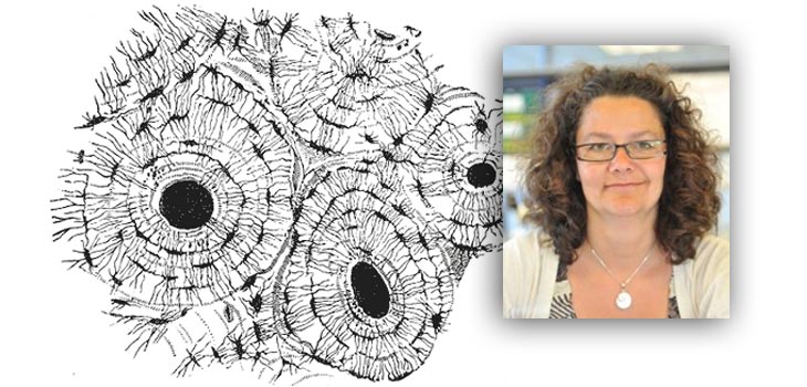
The study was co-led by Dr. Karin Hing, reader in Biomedical Materials at QMUL’s Institute of Bioengineering. As indicated in the December 17, 2015 news release, “By eight to twelve weeks its performance alone matched that of the new graft mixed with the clinical gold standard, called autograft, which is made up of the patients’ own bone containing living cells and growth factors.”
Asked what makes their approach so exciting and unique, Dr. Hing told OTW, “The combination of an optimised chemistry and an optimised pore structure. Regarding the former, the graft is made from a silicate-substituted calcium-phosphate, hydroxyapatite, which has a similar crystal structure to natural bone mineral. We believe that one of the key beneficial effects of this chemical modification is to alter the surface chemistry to aid recruitment and enrichment of key proteins to the surface of the graft, such as adhesion proteins and growth factors which in turn aids bone cell attachment, proliferation and development.
“As for the optimised pore structure, the graft has two ‘levels’ of porosity, macropores (that can be seen by eye) and strut pores (tiny pores within the dense ‘struts’ of the graft). Both of these levels of porosity are highly interconnected to enable cell and blood vessel penetration into the macroporosity and nutrient (and possibly even cell) transfer across the graft struts. This is similar to the sort of multi-level pore structure found in cancellous bone, where in addition to the ‘sponge-like’ macropore structure of cancellous bone, the individual trabeculae of bone contain an interconnected network of micro-pores known as lacunae, within which the osteocyte bone cells reside.”
“Both these factors combine to produce a synthetic bone graft substitute material that is able to support and guide bone’s native regenerative ability to promote bone regeneration at the treatment site.”
“Having developed synthetic bone grafts which perform well in pre-clinical trials and clinical practice (products are available for clinical use through Baxter Inc. who acquired the university spin-out company ApaTech Ltd., that we set up to move our research from the bench to the bedside). The next challenge is to understand how a synthetic ceramic based graft is able to guide/promote osteoproductive behaviour. This knowledge could be used to further develop ‘smart’ synthetic bone grafts and should also be communicated to surgeons so that they can make more educated, confident decisions about the type of bone graft that they want to use in theatre. To this end we are running a number of different research projects looking at how different aspects of the grafts structure and chemistry may affect the biological response.”
“That optimised bone graft substitute materials have the potential to support bone regeneration in situ, by adopting the bodies’ native bone regenerative capabilities. For some patients this may negate the need for a second operative procedure to harvest autograft, so reducing theatre and patient recovery time and the added risks of donor site infection, morbidity and chronic pain.”
React:
Discussion
This is a fascinating development. In my practice we've seen similar outcomes with the revised protocol. The key differentiator seems to be patient selection criteria. Has anyone else noticed the correlation with BMI thresholds?
Great point. I'd push back slightly on the conclusion, the sample size in the cited study is too small to draw population-level inferences. That said, the directional signal is compelling and worth a larger RCT.
We implemented a similar approach last year. Early results are promising but we're still gathering 12-month follow-up data. Happy to share our protocol if anyone is interested.
Join the conversation
Orthopedic professionals are discussing this. Sign in and upgrade to read every comment and add your voice.