Thanks to researchers at the Salk Institute, for the first time it is possible to watch motor neurons—in real time—as patients walk. The work, published in Neuron, is laying the groundwork for a deeper understanding of how spinal cord cells make connections with motor neurons. Such knowledge could aid medical professionals in repairing broken connections in patients with spinal cord injuries or neurodegenerative diseases.
Watch Neurons Walk! Hope for Spinal Cord Injury?
2 min read Premium comments
Secondary
“Using optical methods to be able to watch neuron activity has been a dream over the past decade, ” says Samuel Pfaff, Ph.D., a professor in Salk’s Gene Expression Laboratory, in the September 2, 2015 news release. “Now, it’s one of those rare times when the technology is actually coming together to show you things you hadn’t been able to see before.”
According to the news release, “In the past, to measure the activity of neurons—whether in the brain or extending throughout the body—scientists relied on electrodes that could detect the change in electrical voltage inside a cell when it’s activated. But it is tricky to use electrodes to simultaneously record the activity of many different neuron types at once to study how their activity is synchronized.”
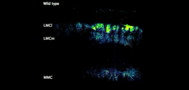
“Pfaff’s team, however, used a fluorescent sensor protein called GCaMP6f that lights up whenever a neuron is activated. Unlike the electrodes, the protein could easily be added to many different cells at once. When Pfaff and his colleagues added GCaMP6f to motor neurons, they could watch with a microscope which cells were activated in a mouse spinal cord when chemicals that turn on walking circuits were added.”
“By tweaking the locations and identities of motor neurons, and then watching the resulting patterns of activation using their new fluorescent technique, Chris Hinckley in the Pfaff laboratory found that the CPG didn’t rely solely on the cells’ locations to connect to them. Instead, the genetic identity of each subtype of cells is also important.”
“Currently, many scientists are attempting to turn stem cells into motor neurons, which they then implant into the spinal cord to regenerate damaged connections. Pfaff’s new results, though, suggest that general motor neurons might not do the trick–the best treatment may require the right subtypes of motor neurons. More work, however, is needed to understand the implications of this and exactly how it might translate to disease treatment.”
Dr. Pfaff told OTW, “We were surprised to find that many aspects of spinal circuitry controlling rhythmic firing of motor neurons associated with stepping were established regardless of motor neuron position within the spinal cord or subtype identity associated with connections to specific muscles. What changed when the identity of motor neurons was perturbed was the coordination of different motor groups.”
Asked what orthopedic surgeons and researchers need to know about this work, Pfaff commented, “Some aspects of motor circuitry are quite robust, namely, the components controlling rhythm. The finer control of muscle coordination is based on a much more specific circuitry.”
React:
Discussion
This is a fascinating development. In my practice we've seen similar outcomes with the revised protocol. The key differentiator seems to be patient selection criteria. Has anyone else noticed the correlation with BMI thresholds?
Great point. I'd push back slightly on the conclusion, the sample size in the cited study is too small to draw population-level inferences. That said, the directional signal is compelling and worth a larger RCT.
We implemented a similar approach last year. Early results are promising but we're still gathering 12-month follow-up data. Happy to share our protocol if anyone is interested.
Join the conversation
Orthopedic professionals are discussing this. Sign in and upgrade to read every comment and add your voice.